가격 및 샘플 지원 요청
지르코니아 크라운, 리튬 디실리케이트 크라운, 베니어, OEM 서비스 또는 도매 수복물 주문을 비교하고 있는 치과 기공소, 치과 진료소, 유통업체 및 조달 담당자분들을 위한 정보입니다.
귀사의 제품 유형, 소재, 월간 생산량, 수출 대상국 및 샘플 요청 사항을 알려주시면, 당사 영업팀이 적절한 후속 조치를 준비할 수 있습니다.

Veneer preparation design is more than a question of how many tenths of a millimeter to remove. The incisal decision (keep the edge, bevel it, or wrap ceramic onto the palatal surface) determines where bonded ceramic is loaded, where the margin sits, how much length the laboratory can add, and what a scanner and technician must be able to read. Window, incisal bevel, and palatal overlap preparations each solve a different problem, and none of them is the right default for every tooth.
This guide defines the three designs, compares them, explains where the clinical and laboratory evidence agrees and where it conflicts, and lists what a dentist should specify before sending a case to a veneer laboratory. It is written for dentists, lab managers, and clinic buyers who order veneers from outside laboratories, and it is a planning resource rather than a substitute for the treating clinician’s diagnosis.
Veneer preparations are usually classified by what they do at the incisal edge. A 2018 critical review of incisal preparation design for ceramic veneers 에서 Journal of the American Dental Association describes four common designs: window (intraenamel), feathered edge, palatal chamfer (overlapped), and butt joint (also called incisal bevel). Window and feathered-edge designs are non-overlap preparations. Butt joint and palatal chamfer are overlap preparations.
In practical terms:
Terminology is not used identically in every publication, so “bevel” or “overlap” can mean different amounts of reduction to different clinicians. To avoid guesswork, a lab prescription should name the design in words and state the intended incisal reduction and palatal finish-line position for each tooth.
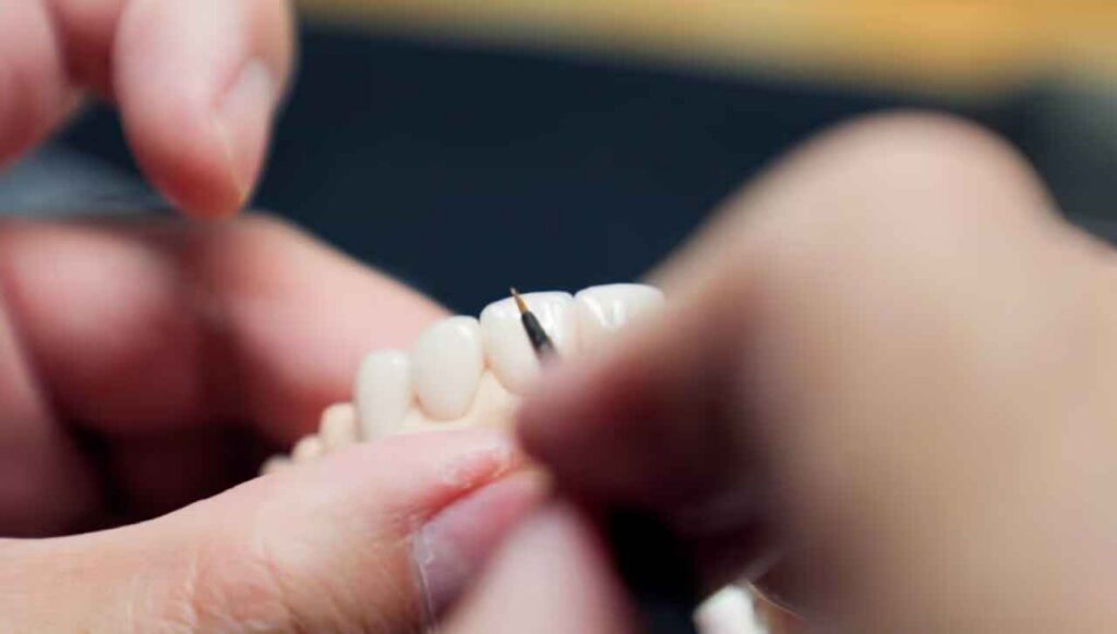
A window preparation keeps the natural incisal edge and bonds ceramic only to the facial surface. Its main advantage is tissue conservation and the possibility of keeping the margin inside enamel.
The enamel argument is supported by data. In a retrospective study of 580 porcelain laminate veneers placed in 66 patients and followed for up to 12 years, overall cumulative survival was 86%. Veneers confined to enamel showed 99% survival, and those with enamel only at the margins showed 94%. Veneers bonded to dentin, or with margins in dentin, were roughly ten times more likely to fail than those bonded to enamel. Artist Dental Lab’s article on how much enamel preservation matters in veneer preparation covers the substrate side of this decision in more detail.
A window design fits best when:
It fits poorly when the plan requires added length, because the extra length would be carried by ceramic that has no tooth structure behind it. It is also a weak choice in edge-to-edge relationships. Artist Dental Lab’s discussion of veneers in deep bite or edge-to-edge cases refers to a finite element analysis that found severe stresses with window preparation under edge-to-edge loading.
Illustrative scenario (hypothetical, not a customer case): a clinic plans window preparations on four worn maxillary incisors and asks for 1 mm of additional length. The laboratory can only add that length as free-standing ceramic beyond the natural edge. A conversation before manufacture, about either an incisal bevel or a revised length target, costs less than a fractured incisal edge at the review appointment.
An incisal bevel reduces the incisal edge so the veneer covers it, without carrying a chamfer onto the palatal surface. The restoration meets the tooth at an incisal butt joint.
This design gives the technician room to build incisal translucency, halo effects, and edge thickness, and it allows moderate length correction. It also avoids a palatal margin in the area where opposing teeth often make contact. The 2018 JADA review concluded that its evidence seems to favor the butt joint over the palatal chamfer, noting that in vitro studies linked palatal chamfer designs with a higher risk of ceramic fracture, while butt joint had the least effect on tooth strength. The review also points out that incisal ceramic is the most common location of ceramic fracture, so the edge deserves specific attention regardless of design.
Buyers should treat that as a lean in the evidence, not a rule. The authors themselves flagged a lack of standardization in the finite element models used in these studies.
Ask the clinician to confirm:
A palatal overlap extends the veneer over the incisal edge and onto the palatal surface, ending in a chamfer. It increases bonded area, gives a clear seating reference, and helps when the incisal edge must move in length or position.
Typical reasons to choose it include:
The trade-offs are real. A palatal overlap removes more enamel, places a margin in a zone that may carry functional contact, and gives the technician a thin ceramic wrap over a corner. Opposing contacts, patient parafunction, and lower-incisor position must be checked before this design is selected. In deep bite cases, the palatal surface may have no clearance for added ceramic without further reduction, which is one reason the occlusion discussion should come before the prescription.
The table below is a qualitative planning aid. It does not give reduction depths, because depth must be set by the planned restoration and the enamel available, not by a fixed number.
| Design | What changes at the incisal edge | Best suited to | 주요 한계 | What the lab needs to know |
|---|---|---|---|---|
| Window | Natural edge retained; facial reduction only | Intact edge, minimal length change, favorable occlusion | Limited length control; added length is unsupported ceramic; poor fit for edge-to-edge loading | Substrate shade, enamel remaining, whether edge translucency is expected from the tooth or ceramic |
| Incisal bevel (butt joint) | Edge shortened and covered; margin at the incisal edge | Moderate length correction, incisal characterization, avoiding a palatal margin | Incisal ceramic is a common fracture location; needs clear incisal clearance | Incisal reduction in mm, lower-incisor contacts, edge thickness expectations |
| Palatal overlap (chamfer) | Ceramic wraps onto the palatal surface with a chamfer margin | Larger length or position changes, weak incisal tooth structure, need for seating reference | More enamel removed; margin may sit in contact zone; thin ceramic over a corner | Palatal finish-line position, functional contacts, parafunction, clearance in protrusion |
| Feathered edge | Preparation reaches edge with no defined finish line | Selected conservative cases | Margin is hard to read and to scan | Photographs and notes defining where the edge should end |

The honest answer is that the research does not crown a winner. Three sources point in different directions:
These findings are not contradictory so much as conditional. Stress models change with load angle, material, and geometry, and clinical survival depends on enamel, occlusion, and operator technique at least as much as on the incisal shape.
A common assumption is that keeping the incisal edge is always the lower-risk choice. It is lower risk for tooth structure, and the enamel data above explain why that matters. But it is not automatically lower risk for the ceramic. A window preparation in an edge-to-edge bite, or one asked to deliver added length, can place the ceramic in a worse mechanical position than a covered edge. Likewise, deeper palatal coverage does not automatically add protection if it removes enamel, thins the ceramic over a corner, or moves the margin into a contact zone.
The defensible position is to choose the least invasive design that still gives the technician enough supported ceramic to reproduce the approved mock-up and survive the patient’s function. That conclusion holds when enamel is available and the bite has been assessed. It does not hold when the diagnosis is incomplete.
The incisal design cannot be chosen in isolation. Three neighboring decisions affect it.
Facial reduction and profile. If the planned veneer adds facial volume to an already prominent tooth, the incisal edge position and lip support change together. Artist Dental Lab’s guide to veneer preparation for protrusive teeth, profile, and lip support explains why the design should start from the approved final contour and why profile and lip support should be tested before preparation, not after cementation.
Cervical and facial depth. Educational protocols often reference around 0.3 mm cervically and 0.5 mm through broader facial areas, as summarized in Artist Dental Lab’s enamel article. Enamel thickness varies by tooth and position, so these figures are starting references. They are not guarantees that the cut stays in enamel.
Interproximal extension. Where a margin sits near the contact affects visibility, contact control, and whether the ceramic must be thickened to hide a transition. The article on interproximal extension for veneers and hiding margins without overcontouring explains how to decide how far a preparation should travel before it gives up enamel without improving the result.
Work backward from the approved restoration, then match the design to the case variables.
| Case variable | Design implication | What to confirm |
|---|---|---|
| Length change planned | Window limits length control; bevel or overlap usually needed | Final incisal edge position from mock-up or wax-up |
| Deep bite or edge-to-edge relationship | Occlusion drives the design; window is a poor default | Bite records, protrusive and lateral contacts, parafunction |
| Intact, sound enamel | Favors conservative designs | Photographs showing remaining enamel and dentin |
| Dark or discolored substrate | Needs enough ceramic thickness; may influence edge treatment | Stump shade for each tooth |
| Weak or worn incisal edge | Coverage may be needed to replace unsupported tooth structure | Edge integrity and existing restorations |
| Prominent tooth or lip-support concern | Preparation must create space, not just add ceramic | Profile photographs and mock-up |
Material also affects the design. Hand-layered feldspathic porcelain suits conservative anterior work where thin, translucent ceramic is the goal. The feldspathic veneer product page lists window, butt-joint, and incisal wrap as design options, recommends conservative preparation, and asks for margin design and reduction notes. Thin ceramic wrapping a sharp corner is vulnerable, so rounded line angles and adequate thickness at the wrap are generally preferred. Monolithic lithium disilicate offers more mechanical reserve, but it does not remove the need for space.
These are common failure modes, drawn from the logic of the designs above rather than from a measured survey:
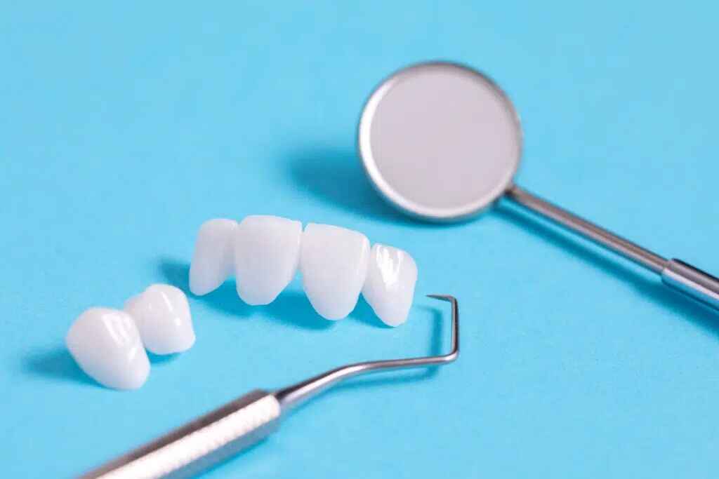
A good preparation can still be lost at the scan. Overlap designs in particular depend on a readable palatal finish line, and thin incisal and interproximal margins can disappear in a poor scan. Artist Dental Lab’s guide to 얇은 마감선을 손상시키지 않고 베니어 준비 부위를 스캔하기 covers margin exposure, moisture control, and how to decide whether to accept or retake a scan.
The laboratory should verify margins independently and report unreadable areas before production starts. A margin the technician has to estimate is a margin that can shift the fit.
A complete prescription removes guesswork. For each tooth, specify the following:
| Information | 중요한 이유 |
|---|---|
| Incisal design (window, bevel, overlap) in words and mm | Terminology varies between clinicians and publications |
| 승인된 왁스업, 모크업 또는 임시 스캔 | Defines final length, contour, and facial position |
| Prepared scan, opposing arch, and bite record | Shows available space and functional contacts |
| Preparation photographs showing enamel and dentin | Tells the technician the bonding substrate and masking demand |
| Stump shade and target shade | Determines ceramic opacity and thickness |
| Margin notes, including the palatal finish line | Prevents the lab from guessing at unreadable areas |
| Parafunction and guidance notes | Influences edge thickness and material choice |
| Material, unit count, and delivery schedule | Affects fabrication workflow and quotation |
Buyers placing wholesale or OEM orders should confirm with the laboratory what file formats it accepts, what happens when a margin cannot be read, and who is responsible for contacting the clinic about thickness conflicts. Do not assume these terms; ask for them in writing.
A window preparation leaves the natural incisal edge intact and bonds ceramic to the facial surface. An incisal overlap covers the edge and may extend onto the palatal surface. Overlap allows more length and edge control but removes more tooth structure.
In the 2018 JADA review, incisal bevel and butt joint are treated as the same design: the incisal edge is reduced and covered without a palatal chamfer. Other publications use the terms less consistently, so prescriptions should state reduction in millimeters.
No design has been shown to be clearly superior. One meta-analysis estimated 88% survival with incisal coverage and 91% without, and reported high survival either way. Enamel availability, occlusion, and technique appear to matter at least as much.
Palatal overlap is considered when the incisal edge must be lengthened or repositioned, or when incisal tooth structure is weak. It requires adequate palatal clearance and a bite assessment, because the margin may sit near functional contacts.
It is generally a poor default. Edge-to-edge and deep bite relationships load the incisal ceramic directly, and occlusion should be diagnosed before any preparation is chosen. Orthodontics, staged treatment, or a different incisal design may be more appropriate.
Preserve as much as the planned restoration allows. In one retrospective study, veneers confined to enamel had 99% survival, versus about ten times higher failure risk with dentin bonding or dentin margins. The target is the least reduction that still gives sufficient ceramic space.
Send the prepared and opposing scans, bite record, approved mock-up or wax-up, preparation photographs, stump and target shades, margin notes, and functional notes. State the incisal design in words and millimeters for each tooth.