للمختبرات السنية والعيادات والموزعين وفرق التوريد التي تقارن بين تيجان الزركونيا وتيجان ثاني أكسيد الليثيوم والقشور وخدمات التصنيع حسب الطلب (OEM) أو طلبات الترميم بالجملة.
يرجى إطلاعنا على نوع المنتج، والمواد المستخدمة، وحجم المبيعات الشهري، وبلد المقصد، وطلب العينات، حتى يتمكن فريق المبيعات لدينا من تحديد الخطوة التالية المناسبة.
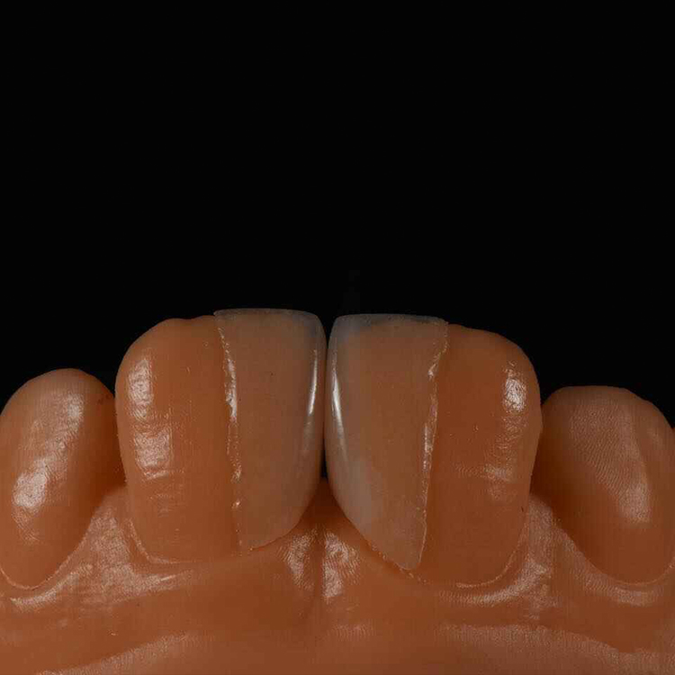
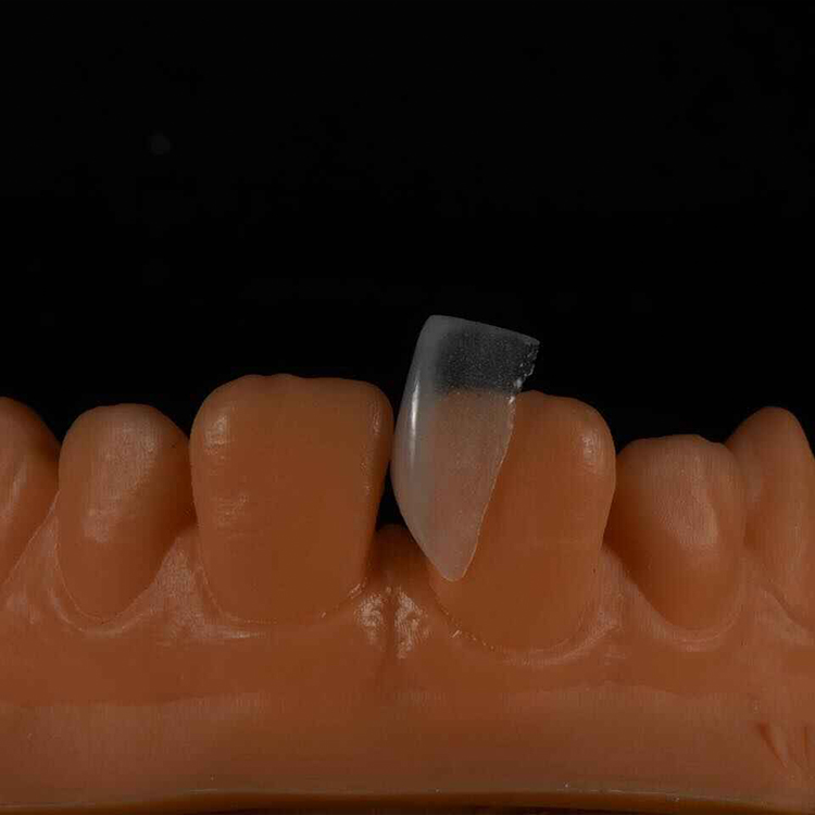
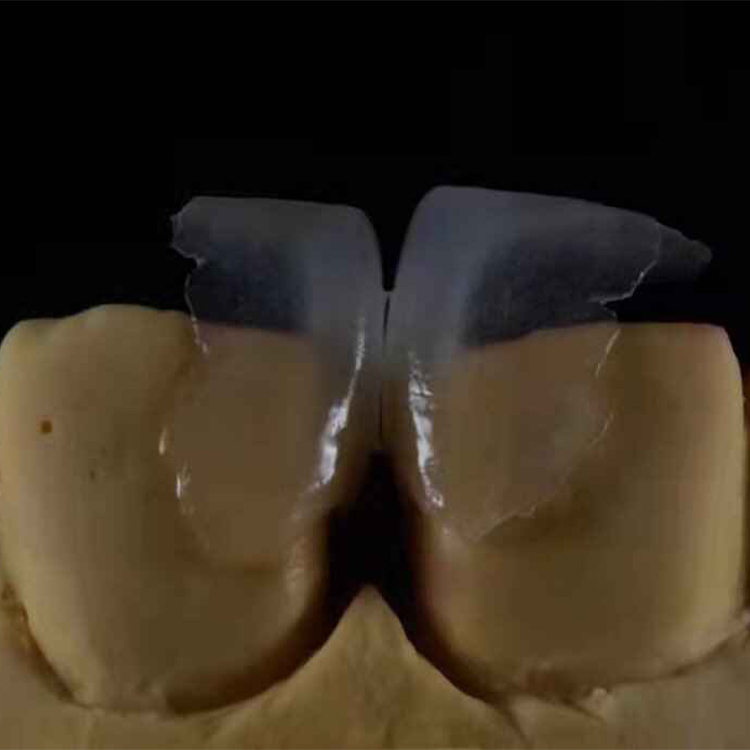
قشور الأسنان ذات الحافة العالية: تصميم السيراميك عند الحد المرئي للثة
لا شيء يبقى خفيًّا.
عندما ترتفع الشفة العليا فوق حواف اللثة، فإن الثلث العنقي، وتناسق الأنسجة، والانتقال إلى السيراميك، وشكل الحليمة، وكل جزء من الكفاف الزائد، تدخل جميعها في مجال رؤية المريض؛ لذا فإن الترميم الذي تم تركيبه بشكل صحيح من الناحية الفنية قد يظل يمثل فشلًا جماليًا واضحًا.
فلماذا، إذن، يوافق العديد من الفرق على تركيب قشور البورسلين بناءً على صورة مقربة مع تراجع اللثة وحدها؟
تبدو عبارة “قشور الأسنان ذات خط الابتسامة العالي” وكأنها تصنيفًا للمواد. لكنها ليست كذلك. فهي تصف حالة ظهور يتم فيها تقييم الترميم والسن والأنسجة اللثوية معًا — غالبًا أثناء الضحك أو التحدث أو تصوير مقاطع الفيديو بالهاتف الذكي، وليس في الصورة الثابتة الخاضعة للرقابة التي تُستخدم أثناء تجربة القشرة.
وفقًا للمراجعة السريرية التي أجرتها المعاهد الوطنية للصحة (NIH) بشأن ظهور اللثة بشكل مفرط, ، حيث يؤدي ارتفاع خط الابتسامة إلى ظهور تيجان الأسنان الفكية بالكامل بالإضافة إلى كمية زائدة من اللثة. وتحدد الدراسة الاستعراضية نفسها عدة أسباب محتملة، منها تغير عملية البروز السلبي، والانزياح السني-اللحمي، والزيادة الرأسية في الفك العلوي، وفرط حركة الشفة العليا.
هذا التمييز مهم. يمكن لقشور البورسلين أن تغير الطول والعرض واللون والملمس والتناسب الظاهري للأسنان. لكنها لا تستطيع تصحيح جميع أسباب «الابتسامة اللثوية».
خط الشفاه هو اختبار للتحمل، وليس علامة تجميلية
إن خط الشفة المرتفع يبرز الاختلافات الطفيفة التي قد يخفيها الموضع العادي للشفة. فلم يعد ارتفاع قمة اللثة بشكل طفيف، أو الانتقال الرمادي عند عنق السن، أو المظهر البارز للسن عند ظهوره مجرد ملاحظة هامشية في المختبر، بل أصبح جزءًا من الابتسامة.
أنا أتبع قاعدة تخطيط واضحة وصريحة: إذا لم يستطع الثلث العنقي الصمود أمام كل من مقطع فيديو يظهر فيه الابتسامة الكاملة وصورة ماكرو بدون إضاءة، فإن التصميم لم يكتمل بعد.
يمكن أن تغير الفينير التناسبات، لا التركيب البيولوجي
غالبًا ما يتلقى المرضى الذين يبحثون عن “قشور الأسنان لعلاج الابتسامة اللثوية” إجابةً مريحةً للغاية: قم بإطالة الأسنان وستبدو اللثة أصغر حجمًا. أحيانًا ينجح ذلك. وأحيانًا يؤدي إلى ظهور أسنان كبيرة الحجم في ظل نفس الظهور المفرط للثة.
يجب على الطبيب المعالج أن يحدد أولاً السبب الكامن وراء ظهور هذه الحالة:
ثوران سلبي متغير
التيجان السريرية القصيرة أو البالية
تضخم اللثة
الانزياح السني-اللحمي
الفائض الرأسي في الفك العلوي
شفة علوية قصيرة أو شديدة المرونة
قمم اللثة غير المتماثلة
مزيج من عاملين أو أكثر
إذا كان حجم الأسنان هو المشكلة الرئيسية، فقد يوفر تصميم الفينير الخزفي حلاً محافظاً. أما إذا كانت المشكلة هيكلية أو عضلية، فإن الفينير وحده لا يعدو كونه إخفاءً تجميليًّا — وغالباً ما يكون إخفاءً غير فعال.
هذه هي الحقيقة المرة. لا يمكن للغلاف الخزفي أن يتكيف مع الفك العلوي.
كيفية تصميم الفينير لخط ابتسامة مرتفع
الصور الثابتة تُظهر الشخص في أفضل حالاته.
يسجل فيديو «الابتسامة الطبيعية» مدى سرعة ارتفاع الشفة، وما إذا كان الجانبان الأيمن والأيسر يتحركان بشكل متماثل، ومدى ظهور اللثة أثناء الضحك التلقائي، وما إذا كان التصميم الذي يبدو مقبولاً يفقد مظهره الجيد بمجرد أن يتوقف المريض عن التمسك بابتسامة تم التمرين عليها في عيادة الأسنان.
ماذا يحدث خارج إطار الصورة؟
التقط الابتسامة وهي تتحرك
بالنسبة لفينير خط الابتسامة العالي، يجب أن تتضمن الحزمة المرسلة إلى المختبر أكثر من مجرد ملف STL ووصفة A1. وأود أن أرى، على الأقل، ما يلي:
صور للوجه في وضع الاسترخاء التام، وابتسامة طبيعية، وابتسامة واسعة
مقطع فيديو مدته من خمس إلى عشر ثوانٍ يُظهر الكلام وحركة الابتسامة العفوية
مناظر أمامية وجانبية مُسحبة
صورة جانبية وصورتان للوجه بزاوية ثلاثة أرباع
صورة فوتوغرافية في وضع الساعة 12 لتحليل المستوى القاطع وخط الوسط الوجهي
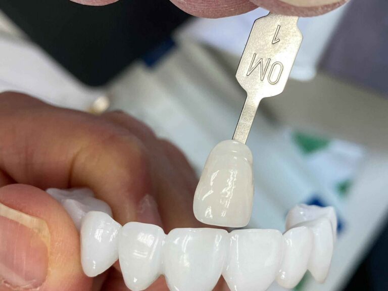
صور لأسنان معالجة مع علامة تحدد لون الجذع
الصور ذات الاستقطاب المتقاطع عندما يصعب قراءة القيمة واللون الداخلي
التصوير الشعاعي للفكين العلوي والسفلي، والعضة الشدقية، والتشريح قبل الجراحة
النموذج الشمعي أو النموذج التجريبي أو المسح الضوئي المؤقت المعتمد
ملاحظات مكتوبة تتناول خط الشفاه، والإطباق، والوظائف غير الطبيعية، والملمس، والشفافية
دليل «مختبر أرتيست للأسنان» حول إرسال حالات الفينير الأمامي إلى المختبر ويوسع نطاق تلك المجموعة من السجلات لتصبح قائمة مرجعية عملية يمكن استخدامها من العيادة إلى المختبر.
كما يحتاج المختبر إلى هدف بصري محدد. فمصطلح “طبيعي” ليس وصفة طبية. فهل يطلب المريض ابتسامة من النوع BL2، أم ابتسامة من النوع A1 تتناسب مع العمر، أم هالة قاطعة مرئية، أم لمعان سطحي منخفض، أم مطابقة دقيقة للأسنان النابية غير المعالجة؟ الإطار المرجعي ذو الصلة لتحديد خمسة أهداف جمالية قابلة للقياس في مجال قشرة الأسنان تفرض اتخاذ تلك القرارات قبل تصنيع السيراميك.
قم أولاً بإنشاء الواجهة ذات اللونين الوردي والأبيض
يبدأ تصميم الابتسامة الجمالية للثة بالنقاط العظمية العلوية للثة، والحليمات، ونسب التاج السريري — وليس باللوحة اللونية النهائية.
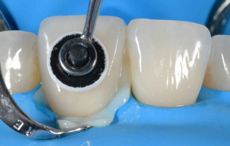
وثّق تقرير حالة من عام 2024 مسار عمل يجمع بين نموذج شمعي رقمي، ونموذج تجريبي داخل الفم، ودليل لاستئصال اللثة مطبوع بتقنية ثلاثية الأبعاد، ودليل لتقليص الأسنان، وقشور خزفية مصنوعة يدويًّا تم لصقها تحت عزل الحاجز المطاطي. وتكمن قيمة ذلك في حالة استئصال اللثة وتركيب الفينير باستخدام التوجيه الرقمي لم تكن مجرد عملية طباعة. فقد تم تخطيط بنية اللثة وخطوط السيراميك استنادًا إلى نفس التصميم المعتمد.
هذا هو النموذج الذي أثق به: التخطيط للأنسجة والسيراميك كنظام بصري واحد، مع إبقاء الإجراءات المتعلقة باللثة والترميم تحت إشراف الأطباء المتخصصين المعنيين.
لكن الجراحة ليست إجراءً تلقائيًّا يسبق تركيب الفينير. فإذا أدى إطالة التاج إلى كشف الجذر، أو إتلاف الدعم الحليمي، أو تسبب في عدم استقرار مستويات الأنسجة، فقد تصبح الابتسامة أقل طبيعية بعد العلاج. لذا، يجب أن يتضمن الخطة الفحص التشخيصي، وتقييم مستوى العظم، وتقييم النمط الظاهري للأنسجة، وفترة الشفاء، قبل البدء في التحضير الذي لا رجعة فيه.
وضع حواف القشرة: توقف عن إخفاء المشاكل تحت اللثة
إن إخفاء الحافة لا يجعلها تختفي. بل يجعل من الصعب فحص المشكلة ومسحها وتثبيتها وتنظيفها وصيانتها.
قد يدفع ارتفاع خط الابتسامة الفريق إلى مد خط النهاية إلى عمق أكبر داخل التجاويف، نظرًا لظهور الحد العنقي. وأعتقد أن هذا التصرّف الغريزي هو السبب وراء عدد لا بأس به من حالات الفينير المصابة بالالتهاب، ذات الحواف الرمادية، والتي يصعب الحفاظ عليها.
كلما سمحت الظروف البصرية والهيكلية بذلك، فإن الحافة فوق اللثة أو المتساوية مع اللثة توفر عزلًا أنظف، وارتباطًا أكثر قابلية للتنبؤ به مع المينا، وسهولة أكبر في أخذ القالب أو إجراء المسح الضوئي، وصيانة أفضل. أما الامتداد تحت اللثة فيجب أن يستجيب لحاجة محددة — مثل إخفاء عيب موجود أو ترميم سابق — وليس بسبب القلق من ظهور الحواف.
التجويف ليس مساحة تخزين
يختلف الارتباط النسيجي فوق الحافة السنية — الذي كان يُعرف سابقًا باسم “العرض البيولوجي” — من مريض لآخر ومن منطقة لأخرى. واعتباره «قاعدة عامة» ثابتة تبلغ «3 ملم» هو ممارسة طب أسنان متهاونة.
مراجعة سريرية صدرت عام 2024 حول انتهاكات ارتباط الأنسجة فوق الحافة السنية يصف العواقب المحتملة للتعدي على الترميم، بما في ذلك الالتهاب، وتراجع اللثة، وتشكل الجيوب، وفقدان الارتباط. وفي حالة المريض الذي يعاني من ارتفاع خط الشفة، لا يمثل تراجع اللثة مجرد ضرر بيولوجي فحسب، بل يمكن أن يكشف عن حافة القشرة التي حاول الفريق إخفاءها.
«جينجيفا» تسجل النتائج.
عندما يتم بناء محيط عنق السن بشكل مفرط لإخفاء جذع الأسنان الداكن، أو عندما يتم تحديد خط النهاية باتجاه القمة دون إجراء تقييم كافٍ للأنسجة اللثوية، فقد تظهر الترميمات بشكل جيد في الصور في البداية، لكنها تصبح معيبة بشكل واضح بعد نضوج الأنسجة وتأثيرات التحديات الروتينية المتعلقة بنظافة الفم.
هل كان الهامش الخفي يستحق العناء؟
السيطرة على ظهور العناصر قبل إضافة التوصيف
عادةً ما يتطلب الثلث العنقي المقنع ما يلي:
انتقال سلس من سطح الجذر إلى السيراميك
لا يُسمح باستخدام السيراميك غير المدعوم أو المتدلي
قمم اللثة متماثلة ولكنها ليست متطابقة ميكانيكيًا
خطوط انحناء بين الأسنان تدعم الحليمات دون الضغط عليها
نسيج سطحي يصبح أكثر نعومة كلما اقترب من منطقة عنق الرحم
التحكم في الفلورة والقيمة والعتامة فوق الجذع
حافة يمكن للطبيب عزلها وإنجازها
قد يكون خطأ في محيط السطح الأمامي العنقي يبلغ 0.2 ملم أكثر إضرارًا بالمظهر النهائي من اختلاف طفيف في شفافية الحافة القاطعة. ومع ذلك، يُطلب من المختبرات بشكل روتيني مناقشة مسألة الهالات في حين أن ملامح ظهور السن لا تزال دون حل.
هذا تفكير رجعي.
اختيار السيراميك عند الحد الظاهر للثة
لا أعتبر تحديد نوع المادة دليلاً على التشخيص.
يمكن أن ينتج كل من البورسلين الفلسباتي، وثاني سيليكات الليثيوم، والزركونيا الشفافة ترميمات يمكن الدفاع عنها. لكنها لا توفر نفس خصائص التغطية، أو طريقة الالتصاق، أو التفاوت المسموح به في السماكة، أو استجابة العنق للضوء. مختبر «أرتيست دنتال» دليل مواد الفينير الأمامي القائم على الحالات السريرية يوفر منطق الاختيار الأوسع نطاقاً.
المواد
الكيمياء والنظام
أفضل استخدام لـ«خط الابتسامة العالي»
مخاطر سرطان عنق الرحم
اعتبارات الترابط
خزف فلدسباثي
سيراميك مصنوع من السيليكا، مُركَّب يدويًّا بطبقات
حالات تتسم بغزارة المينا مع تغير طفيف في اللون ومتطلبات بصرية عالية جدًّا
شفافية زائدة فوق الجذوع الداكنة؛ حواف رقيقة تتأثر بالتقنية المستخدمة
يتم معالجتها عادةً بالهيدروفلوريد (HF) والسيلان وفقًا لتعليمات الشركة المصنعة للسيراميك
ثنائي سيليكات الليثيوم
Li₂Si₂O₅؛ يُستخدم عادةً مع أنظمة IPS e.max
قشور البورسلين متعددة الوحدات التي تتطلب توازنًا بين الشفافية والتغطية وقابلية التكرار
انتقال عنقي رمادي أو عالي القيمة في حالة تجاهل لون الجذع والأسمنت
عادةً ما يتضمن بروتوكول الزجاج الخزفي استخدام حمض الهيدروفلوريك والسيلان؛ أما التوقيت فيختلف باختلاف المنتج
ثنائي سيليكات الليثيوم متعدد الطبقات
لب من مادة Li₂Si₂O₅ مع طبقة خارجية من السيراميك
الحالات الأمامية عالية التفاصيل التي تتطلب عمقًا داخليًّا وتأثيرات قاطعة مخصصة
زيادة حساسية التقنية واحتمال حدوث تشققات في المناطق الرقيقة أو التي تتحمل أحمالاً
يعتمد الإجراء على سطح الترابط المكشوف وعلى تعليمات الشركة المصنعة
الزركونيا الشفافة
ZrO₂
حالات مختارة تتطلب قدرًا أكبر من التغطية أو الجهد الوظيفي
العتامة الزائدة، وصعوبة محاكاة المينا، وظهور قفزة ملحوظة في اللون عند منطقة العنق
لا يُعد HF فعالاً على الزركونيا؛ ويُعتبر الكشط الهوائي والأنظمة التي تحتوي على MDP من الخيارات الشائعة وفقاً لبروتوكول الشركة المصنعة
غالبًا ما يكون ثنائي سيليكات الليثيوم خياري الأول في حالات الأسنان متعددة الوحدات ذات خط الابتسامة المرتفع، لأنه يمثل الحل الوسط المثالي: سيراميك زجاجي قابل للربط، وشفافية قابلة للتحكم، وقابلية تكرار أعلى مقارنة بالأعمال الفلسباتية الراقية المُركبة يدويًّا. الـ سير عمل قشرة القشرة E.max ويكون ذلك مهمًا بشكل خاص عندما تتمكن العيادة من توفير صورة لجزء الجذر المتبقي، وحواف واضحة، وصور للابتسامة، ومرجع جمالي معتمد.
لكن كلمة “غالبًا” لا تعني “دائمًا”.”
قد يكون البورسلين الفلسباتي هو الخيار الأمثل عندما يكون المينا وفيرًا، وتكون التغيرات في اللون طفيفة، وتتطلب الحالة تأثيرات سطحية وداخلية دقيقة. أما الزركونيا فقد تدخل في الاعتبار عندما تكون مخاطر التغطية أو المخاطر الوظيفية شديدة. ما أرفضه هو اختيار ZrO₂ لمجرد أن معدل قوته يبدو مثيرًا للإعجاب في الكتيب الترويجي.
لا تفشل الخزف بمفرده. بل تفشل معه عملية اختيار القالب، والتحضير، والربط، والإطباق، والتواصل.
ماذا تقول بيانات البقاء على قيد الحياة فعليًّا؟
الأرقام الأخيرة تبعث على الاطمئنان، لكنها لا تعني أنه يمكننا الاستعداد بشكل مكثف.
أفاد تحليل تلوي مُدرج في قاعدة بيانات «بيوميد» (PubMed) ونُشر عبر الإنترنت في عام 2024 أن معدلات البقاء على قيد الحياة المجمعة بعد مرور 10.4 سنوات بلغت 96.131.13% للقشرة الفلسباتية و 96.81% لقشور ثنائي سيليكات الليثيوم. بلغت نسبة السيراميك الزجاجي المقوى بالليوسيت 93.70%. ولم يجد المؤلفون أي فرق ذي دلالة إحصائية في معدل البقاء بين تلك المجموعات من السيراميك، على الرغم من تباين أنماط المضاعفات.
والآن، لننتقل إلى الإحصائية الأكثر إثارة للقلق.
تلا ذلك تحليل رجعي لعام 2025 672 قشرة خزفية لمدة تتراوح بين سنة واحدة و15 سنة. سُجلت حالات الفينير الملصقة بالكامل على المينا 99% البقاء, ، مقارنةً بـ 94% عندما كانت الطبقة الملتصقة تتضمن كلًّا من المينا والعاج. الـ دراسة حول تعرض العاج ومدى بقاء الفينير يُسلط الضوء على نقطة تميل صناعة مستحضرات التجميل إلى إخفائها: فقد يكون الحفاظ على المينا أكثر أهمية من تحسين مظهر السطح الخزفي.
قد تبدو خمس نقاط مئوية نسبة ضئيلة، إلى أن يتم ضربها في ثماني أو عشر وحدات أمامية، وعدة سنوات من الخدمة، وعمليات إعادة التصنيع، ووقت العلاج على كرسي الأسنان، ومريضة تلاحظ كل شيء لأن حافة لثتها ظاهرة بالكامل.
البيانات تتعلق بالشؤون الشخصية.
لذا، فإن أفضل الفينير لعلاج «الابتسامة اللثوية» ليست بالضرورة الأكثر بياضًا أو الأكثر متانة أو الأغلى ثمناً. بل هي عمليات الترميم التي تعالج مشكلة نسب الأسنان التي تم تشخيصها بشكل صحيح، مع الحفاظ على أفضل سطح ربط ممكن ومراعاة الغلاف النسيجي.
مسار العمل الذي يقلل من حالات إعادة العمل في منطقة خط الابتسامة العالي
1. التشخيص قبل التصميم
حدد ما إذا كانت المظاهر السريرية تتعلق بشكل أساسي بالأسنان، أو اللثة، أو الهيكل العظمي، أو العضلات، أو مختلطة. قم بتسجيل حالة صحة اللثة، والنمط الظاهري للثة، وطول التاج السريري، والعلاقة بين خط التماس بين اللثة والسن (CEJ) والعظم، وموضع الأسنان، وحركة الشفاه.
2. اعتمد الابتسامة بشكل ديناميكي
استخدم نموذجًا شمعيًّا ونموذجًا تجريبيًّا داخل الفم. تحقق من التصميم في وضع الراحة، وأثناء الكلام، وعند الابتسامة الطبيعية، وأثناء الابتسامة القصوى. إن الموافقة على المنظر عند سحب الشفاه فقط هي الطريقة التي تسمح للقشرة الفموية كبيرة الحجم بالمرور بنجاح خلال اجتماع التخطيط.
3. تحديد نقاط النهاية للأنسجة قبل التحضير النهائي
في حالة الحاجة إلى علاج اللثة، حدد النقاط القصوى المستهدفة واترك للطبيب المعالج تحديد الإجراء المناسب وفترة الشفاء. لا تجعل الفني يخمن المكان الذي قد تستقر فيه اللثة في النهاية.
4. الحفاظ على المينا بشكل متعمد
استخدم النموذج المعتمد أو دليل التخفيض المراقب لتوجيه عملية التحضير. يجب أن يتناسب التخفيض مع الحجم المطلوب للترميم، وليس أن يكون قطعًا آليًّا بعمق موحد على جميع الأسنان.
كلما زاد التخفيض، زادت المساحة المتاحة. كما أنه يؤدي إلى تآكل المينا.
5. إرسال وصفة طبية للنظارات
توثيق اللون النهائي، ولون الجذع، وسماكة السيراميك، والقيمة المطلوبة، ومناطق الشفافية، وتأثيرات الحافة القاطعة، وملمس السطح، واستراتيجية استخدام الراتنج واللاصق. لا يقوم مركب Li₂Si₂O₅ بمفرده بإزالة التحضير الداكن.
6. فحص الثلث العنقي عند تجربة الملابس
قم بتقييم الترميمات بعد ترطيبها ومن مسافة مناسبة للمحادثة. افحص ملاءمة الحواف، وانتقال الألوان، والضغط على اللثة، وظهور الأسنان، ودعم الحليمات، والتناسق عند الابتسامة الكاملة — وليس فقط تحت المباعدات.
7. اتبع بروتوكول الترابط الخاص بكل مادة
لا يمكن استبدال خطوات HF، والسيلان، والتآكل بالجسيمات العالقة في الهواء، و10-MDP ببعضها البعض. ويخضع البروتوكول النهائي لتعليمات الشركة المصنعة للسيراميك، وتعليمات نظام المواد اللاصقة، وشروط العزل، وتقدير الطبيب المعالج.
الأسئلة الشائعة
ما هي قشور الأسنان ذات خط الابتسامة العالي؟
قشور الأسنان ذات خط الابتسامة العالي هي ترميمات رقائقية من البورسلين أو السيراميك، مصممة للمرضى الذين تكشف ابتسامتهم الكاملة عن التاج السريري الفكي العلوي بالكامل بالإضافة إلى شريط مرئي من اللثة، مما يجعل محيط عنق السن، وموضع الحافة، وتناسق القمة، ولون الجذع، واستقرار الأنسجة أكثر وضوحًا مقارنةً بحالات خط الابتسامة العادي.
وهي تتطلب سجلات ديناميكية للوجه ورقابة أكثر صرامة على المنطقة الفاصلة بين اللثة والأسنان. ويشير مصطلح “خط الابتسامة المرتفع” إلى نمط عرض المريض، وليس إلى مادة قشرة منفصلة أو تصميم تحضيري معين.
هل يمكن للفينير أن يعالج «الابتسامة اللثوية»؟
لا يمكن للفينير أن يحسّن «الابتسامة اللثوية» إلا عندما تشكل الأسنان القصيرة أو المتآكلة أو الصغيرة الحجم أو غير المتناسقة جزءًا رئيسيًّا من المشكلة البصرية؛ فهي لا تصحح بشكل مباشر الفائض الرأسي في الفك العلوي، أو فرط حركة الشفة العليا، أو بروز الأسنان واللثة، أو النسيج اللثوي الزائد الذي يتطلب علاجًا في مجال أمراض اللثة أو تقويم الأسنان أو الجراحة.
يجب تشخيص السبب قبل البدء في العلاج. فالقشور التي تكتفي بإطالة الأسنان المتناسقة أصلاً قد تجعل الابتسامة تبدو ثقيلة وأقل طبيعية، كما تجعل الحفاظ عليها أكثر صعوبة.
أين ينبغي وضع حواف القشرة في حالة خط الابتسامة المرتفع؟
ينبغي وضع حافة القشرة في حالة خط الابتسامة المرتفع في الموضع الذي يسهل تنظيفه، ويكون قابلاً للالتصاق، ومتوافقاً مع الأنسجة، مع استيفاء المتطلبات البصرية في الوقت نفسه؛ وعادةً ما يُفضل وضعها فوق اللثة أو على مستوى اللثة، إذا أمكن ذلك، مع الاحتفاظ بالامتداد تحت اللثة لتلبية احتياجات محددة تتعلق بالإخفاء أو العيوب أو الحواف الموجودة، وفقاً لما يحدده الطبيب المعالج.
يُعد الهامش الظاهر مشكلة تتعلق بالتصميم والتحكم في الألوان. ولا ينبغي أبدًا أن يكون تحريكه نحو القمة بديلاً عن الاختيار الصحيح للسيراميك، أو إدارة لون الجذع، أو محيط العنق.
ما هو أفضل نوع من السيراميك المستخدم في تصنيع الفينير ذات خط الابتسامة المرتفع؟
أفضل نوع من السيراميك لتركيب قشرة ذات خط ابتسامة مرتفع هو الذي يتوافق مع كمية المينا المتوفرة، ولون الجذع، والتغطية المطلوبة، وسماكة الترميم، والحمل الإطباقي، والهدف البصري؛ وغالبًا ما يوفر ثاني سيليكات الليثيوم التوازن الأوسع نطاقًا، بينما يُفضل البورسلين الفلسباتي في الحالات عالية الجمالية التي تتميز بوفرة المينا، أما الزركونيا فتظل خيارًا انتقائيًا وليس تلقائيًا.
في العديد من حالات التركيبات المتعددة الوحدات، يوفر ثنائي سيليكات الليثيوم قوامًا مفيدًا. وقد يصل البورسلين الفلسباتي إلى مستوى بصري أعلى في أيدي الخبراء، في حين أن ZrO₂ ينبغي أن يلبي متطلبات التغطية أو المتطلبات الوظيفية الفعلية.
كيف يتم تصميم الفينير في حالة خط الابتسامة المرتفع؟
يبدأ تصميم الفينير لخط ابتسامة مرتفع بالتشخيص المسببي والتسجيلات الديناميكية للوجه، ثم يتم تحديد قمم اللثة، وموضع الحافة القاطعة، ونسب العرض إلى الطول، وملف الظهور، وموقع الحافة، واستراتيجية لون الجذع، والمواد المستخدمة، والنموذج التجريبي المعتمد، وذلك قبل إجراء أي تحضير لا رجعة فيه للأسنان أو جراحة للأنسجة.
يجب اختبار التصميم النهائي أثناء التحدث والابتسام الطبيعي والابتسام بأقصى درجة. وتُثبت الصورة الملتقطة من مسافة بعيدة تناسق الأسنان؛ لكنها لا تثبت أن الترميم يتناسب مع ملامح وجه المريض.
ما هي أفضل قشور الأسنان القشرية للابتسامة اللثوية؟
أفضل أنواع الفينير لعلاج «الابتسامة اللثوية» هي الترميمات الخزفية التي يتم تحضيرها بطريقة محافظة، ولا يتم اختيارها إلا عندما يكون لحجم الأسنان أو تآكلها أو شكلها أو نسبها تأثير كبير على مظهر اللثة، مع اختيار المواد بناءً على المينا المتبقية، ولون جذع السن، ومستوى التغطية المطلوب، ومدى ظهور الشفاه، والإطباق، وتصميم الابتسامة الذي يوافق عليه المريض.
قد تكون كل من قشور ثاني أكسيد الليثيوم أو قشور الفلسبات مناسبة. وإذا كان السبب الرئيسي يتعلق بالهيكل العظمي أو العضلي أو تقويم الأسنان أو اللثة، فقد يكون الحل الأمثل هو العلاج متعدد التخصصات — أو عدم استخدام القشور على الإطلاق.
أرسل حالة تتميز بخط ابتسامة عريض تستحق التصنيع
لا ترسل إلى المختبر حالة ذات خط شفاه مرتفع مع صورة واحدة للابتسامة، ولون عام، وتعليمات بـ“جعلها تبدو طبيعية”.”
يرجى إرسال جميع الأدلة التالية: ملفات STL المعدة وما قبل الجراحة، والقوس المقابل، وصور الإطباق التي تم التحقق منها، وصور الوجه في وضع الراحة والابتسامة القصوى، والفيديو الديناميكي، وألوان جذوع الأسنان، وملاحظات الحواف، ونقاط نهاية اللثة، ومخاطر الإطباق، والنموذج التجريبي المعتمد، والهدف البصري المكتوب.
ثم اطرح الأسئلة التي تثير الحيرة. هل محيط عنق السن واضح؟ هل تم الحفاظ على المينا؟ هل يمكن إخفاء الجذع دون الإفراط في بناء الطبقة الخزفية؟ هل سيظل التصميم يبدو طبيعيًا عندما يضحك المريض؟