À l'intention des laboratoires dentaires, des cliniques, des distributeurs et des équipes d'approvisionnement qui comparent les couronnes en zircone, les couronnes en disilicate de lithium, les facettes, les services OEM ou les commandes de restaurations en gros.
Indiquez-nous le type de produit, le matériau, le volume mensuel, le pays de destination et votre demande d'échantillon afin que notre équipe commerciale puisse vous proposer la prochaine étape la plus adaptée.
Pourquoi la qualité des résultats varie-t-elle autant d'une clinique à l'autre alors qu'elles utilisent toutes le même scanner ?
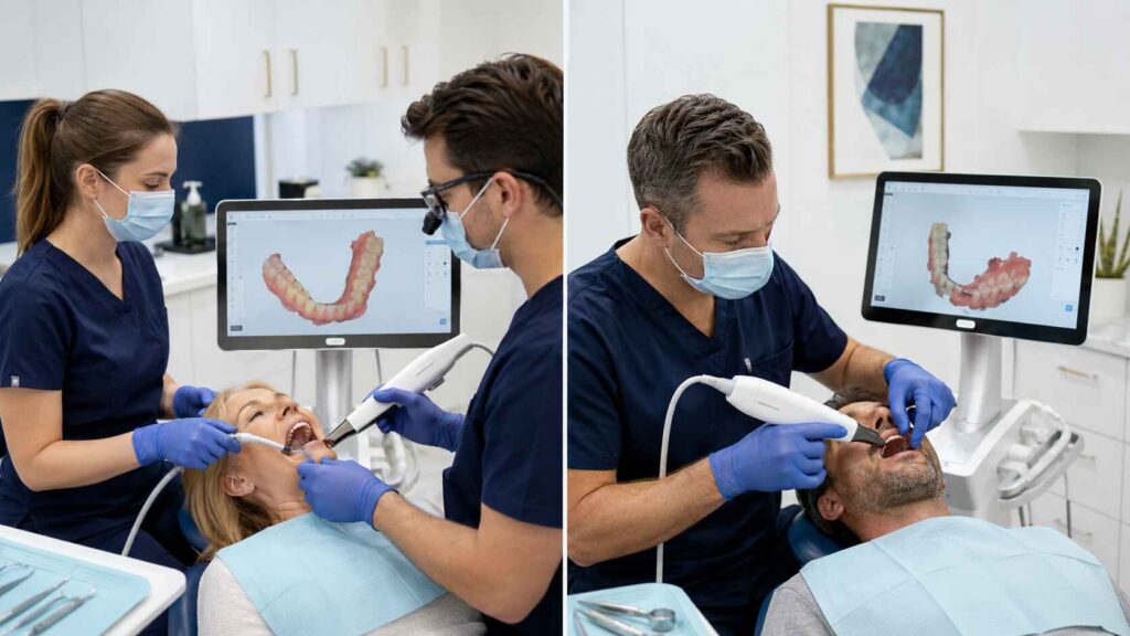
La qualité des cas varie d'une clinique à l'autre, même lorsque celles-ci utilisent le même scanner, car ce dernier ne contrôle pas l'isolation des tissus, le tracé de numérisation, la géométrie de la préparation, les décisions relatives à une nouvelle numérisation, l'enregistrement de l'occlusion, la vérification des fichiers ni les informations transmises au laboratoire.
Le matériel n'y est pour rien.
Un scanner peut certes capturer des millions de points de surface à une vitesse impressionnante, mais lorsqu’un cabinet prend soin de bien définir chaque ligne de finition, suit une séquence de numérisation reproductible, vérifie l’occlusion et rejette les données douteuses, tandis qu’un autre cabinet se contente de numériser à la va-vite la salive, le sang, les tissus en mouvement et une anatomie distale incomplète, les fichiers obtenus sont loin d’être équivalents.
Alors pourquoi les cliniques continuent-elles à rejeter la faute sur le scanner en premier lieu ?
À mon sens, les scanners intra-oraux sont souvent présentés comme des solutions de qualité, alors qu’il s’agit en réalité d’instruments de saisie de données. Ils permettent d’enregistrer ce que l’opérateur leur présente. Ils ne peuvent ni réparer une marge cachée, ni stabiliser une langue qui bouge, ni corriger une préparation présentant une contre-dépouille, ni déterminer si un scan occlusal a été réalisé alors que le patient glissait vers l’intercuspidation maximale.
Le scanner n'est qu'un élément parmi d'autres du processus de travail avec un scanner intra-oral. La restauration finale est le résultat de toutes les décisions cliniques et de laboratoire prises après avoir appuyé sur le bouton d'allumage.
Table des matières
Le scanner n'est pas le système de contrôle qualité
La précision des scanners intra-oraux est généralement évaluée à l'aide de deux paramètres : la justesse et la précision.
La fidélité décrit dans quelle mesure le scan reflète fidèlement l'anatomie réelle du patient. La précision décrit dans quelle mesure le scanner reproduit de manière constante le même résultat lors de scans répétés.
Une clinique peut produire des scans hautement reproductibles qui s'avèrent systématiquement erronés. Elle peut également réaliser une préparation géométriquement acceptable tout en soumettant un cas de restauration de mauvaise qualité, car les marges sont masquées, l'occlusion est instable, l'arcade opposée est incomplète ou encore parce que la prescription adressée au laboratoire se limite à indiquer “ s'harmoniser avec la dent adjacente ”.”
Cette distinction est souvent négligée.
Le logiciel peut afficher un modèle tridimensionnel impeccable. Les couleurs sont attrayantes. La préparation pivote en douceur à l'écran. Pourtant, le fichier peut comporter une gencive « cousue », des surfaces en double, des données proximales manquantes, un bord incorrect, une anatomie postérieure déformée ou une relation occlusale qui ne correspond pas à celle du patient.
Un indicateur de fin de traitement vert ne vaut pas une autorisation clinique.
Les données récentes les plus probantes concernant l'expérience des utilisateurs plaident en faveur d'une position plus nuancée que celle généralement défendue tant par les fabricants de scanners que par leurs détracteurs. A 2025 Journal of Prosthetic Dentistry : revue systématique et méta-analyse a révélé que, dans le cadre de huit études in vitro portant sur 720 scans, la précision des opérateurs inexpérimentés n'était inférieure que de 2,51 μm à celle des opérateurs expérimentés. Cette différence géométrique était minime.
Mais cette même étude a révélé que les opérateurs inexpérimentés avaient besoin en moyenne de 40,95 secondes supplémentaires, et que la satisfaction des patients était inférieure de 12,69%. Après la formation, la précision s'est améliorée de 19,22 μm et la durée du balayage a diminué de 76,46 secondes.
C'est là la véritable leçon à retenir : l'expérience de l'opérateur ne nuit pas toujours à la précision, mais elle influe sur l'efficacité, la confiance, la prise en charge des patients et la capacité à détecter les données erronées avant leur transmission.
Les points faibles réels de la qualité des scans intra-oraux
La précision d'une empreinte numérique ne se joue pas en un seul instant décisif. Elle est généralement compromise par une succession de petits compromis.
Étape du flux de travail
Clinique contrôlée
Une clinique incohérente
Conséquence probable en laboratoire
Gestion des tissus
Ligne d'arrivée visible, hémorragie maîtrisée, terrain sec
Trajectoire normalisée avec une distance constante par rapport au scanner
Mouvements aléatoires, changements fréquents de direction, perte de suivi
Erreurs de couture et déformation de la surface
Nouvelle numérisation
Données erronées supprimées avant leur récupération
De nouvelles données superposées à plusieurs reprises sur des surfaces douteuses
Double anatomie et maillages bruités
Conception de la préparation
Une ligne d'arrivée nette et lisible, avec une réduction adéquate
Transitions brusques, marges profondes non soutenues, jeu limité
Risque d'ajustement et conception de restauration inadéquate
Arc opposé
Anatomie complète et pertinente prise en compte
Dents distales ou surfaces fonctionnelles omises
Conception occlusale peu fiable
Enregistrement des morsures
Données bilatérales vérifiées cliniquement
Une brève analyse de données a été acceptée sans vérification
Occlusion importante ou articulation décalée
Documentation relative à l'affaire
Ombre, ombre projetée par une souche, photos, matériel et instructions de réalisation inclus
Fichiers numérisés envoyés avec une ordonnance imprécise
Une restauration techniquement correcte mais cliniquement inadaptée
Contrôle final
Vérification des marges, des contacts, des trous et de l'occlusion avant l'envoi
Fichier téléchargé immédiatement après la numérisation
Clarification, retard ou refonte évitables
La dure réalité est simple : deux cliniques peuvent utiliser le même scanner TRIOS, Medit, Primescan, iTero ou tout autre modèle et obtenir des jeux de données très différents, car le scanner ne perçoit que le champ optique qui lui est présenté.
Il ne sait pas si ce tissu devrait se trouver à cet endroit.
Il ne sait pas si une surface brillante recouverte de salive masque le bord d'une obturation. Il ne sait pas si le patient a fermé la bouche jusqu'à l'intercuspidation maximale habituelle ou s'il s'est arrêté prématurément parce que l'embout du scanner se trouvait encore entre ses joues.
Et il ne sait certainement pas si la dent a été préparée en laissant un espace suffisant pour la céramique.
L'expérience des opérateurs est importante, mais pas de la manière dont la plupart des équipes commerciales la présentent
L'expression “ dépendant de l'opérateur ” est souvent utilisée à la légère. Elle peut faire référence à la vitesse, au contrôle de la trajectoire de balayage, à la prise en charge du patient, à l'examen des données, à la gestion des tissus mous ou à la précision géométrique. Or, ces éléments ne correspondent pas tous au même résultat.
A Essai clinique prospectif de 2023 Des scans réalisés avec les appareils TRIOS 3 et Medit i500 ont été testés chez 20 patients en orthodontie. Les opérateurs ont été répartis en trois groupes selon leur niveau d'expérience : élevé, modéré et faible.
Dans cette étude, l'opérateur le moins expérimenté a mis nettement plus de temps, et l'i500 a nécessité un temps de numérisation plus long que le TRIOS 3. Cependant, l'expérience de l'opérateur n'a pas eu d'incidence significative sur la précision mesurée entre les canines et entre les molaires. Les chercheurs ont estimé que les opérateurs ayant déjà réalisé environ 50 à 100 numérisations étaient en mesure d'effectuer la numérisation de manière efficace.
Cela ne signifie pas pour autant que la formation soit facultative.
Cela signifie que l'avantage que procure l'expérience se manifeste souvent dans le jugement avant même d'apparaître dans une simple mesure linéaire.
Un opérateur expérimenté est plus à même de reconnaître :
Lorsque le signal a été perdu
Lorsque la salive réfléchissante altère la surface
Lorsqu'un rebord nécessite une rétraction supplémentaire
Quand une zone réanalysée doit être supprimée plutôt que placée en calque
Lorsque la molaire distale n'a pas été entièrement capturée
Lorsque l'analyse de l'occlusion ne correspond pas à l'occlusion visible
Quand un cas d'arcade complète ne doit pas être traité comme une couronne isolée
Quand faut-il interrompre le traitement du dossier avant qu'il n'arrive au laboratoire ?
Les meilleurs opérateurs ne se contentent pas d'être plus rapides. Ils sont plus efficaces pour rejeter les données erronées.
C'est une compétence sous-estimée.
La longueur du balayage et la complexité du cas modifient les calculs mathématiques
Une couronne postérieure isolée, un cas antérieur à six unités et une restauration implantaire sur toute l'arcade ne posent pas les mêmes exigences à un scanner intra-oral.
Les balayages plus courts fournissent des repères anatomiques proches qui aident le logiciel à aligner les images consécutives. À mesure que la longueur du balayage augmente, le logiciel doit relier davantage de segments d'image sur une plus grande distance. De petites erreurs d'alignement peuvent alors s'accumuler.
Les cas d'implantation sur arcade complète édentée sont particulièrement délicats, car il peut y avoir moins de surfaces dentaires distinctives disponibles comme repères de référence. L'angulation des implants, la conception du corps de balayage, la distance entre les implants, la muqueuse mobile, le sang, la salive et la stratégie de balayage peuvent tous influencer le résultat.
Ce degré d'hétérogénéité n'est pas un détail statistique mineur. Il nous indique que les scanners, les protocoles, les configurations des implants, les méthodes de mesure, les opérateurs et les conditions cliniques donnaient lieu à des résultats sensiblement différents.
Cette même revue faisait état de résultats cliniques contradictoires. Une étude a évalué 30 armatures — 15 conventionnelles et 15 réalisées à partir d’empreintes numériques TRIOS — et n’a constaté aucune différence significative en termes d’ajustement. Une autre étude a comparé 50 prothèses conventionnelles et 50 prothèses numériques ; toutes ont été jugées cliniquement acceptables, avec de meilleurs résultats dans le groupe numérique. Un autre essai randomisé a toutefois mis en évidence des écarts plus importants et davantage d’ajustements inadéquats des armatures dans le groupe numérique.
Même catégorie technologique. Des résultats différents.
C'est exactement ça.
La question n'est pas de savoir si les scanners intra-oraux fonctionnent. C'est clairement le cas. La question est de savoir si un cabinet donné a mis en place un protocole de numérisation et de vérification adapté au type de cas soumis.
La préparation et la qualité des tissus restent déterminantes pour ce que le scanner peut détecter
Il existe un mythe tenace dans le domaine de la dentisterie numérique selon lequel une meilleure qualité optique permettrait de compenser une préparation clinique moins soignée.
Ils ne peuvent pas.
Un scanner ne peut pas enregistrer avec précision une ligne de finition qui reste recouverte de tissu. Il ne peut pas créer d'espace de restauration là où la préparation n'en prévoit pas. Il ne peut pas faire la distinction entre un véritable bord sous-gingival et une surface présentant des artefacts dus au sang, à la salive ou à un fil de rétraction en mouvement.
Imaginons deux cabinets dentaires qui numérisent la même préparation de couronne en disilicate de lithium à l'aide du même appareil.
La clinique A maîtrise le saignement, met en place une rétraction adéquate, assèche le champ opératoire, expose toute la circonférence de la marge et inspecte la préparation à fort grossissement avant de transmettre le dossier.
La clinique B réalise l'examen immédiatement après la préparation, alors que le tissu est enflammé et que la marge distale reste partiellement masquée.
Les deux fichiers devraient s'ouvrir sans problème.
Un seul d'entre eux fournit au technicien des informations fiables.
Le même problème se pose dans le cas des facettes. Un beau scan facial ne suffit pas à compenser une anatomie palatine incomplète, des bords proximaux flous, l'absence de dentition antagoniste ou une occlusion imprécise. Artist Dental Lab’s Guide de soumission des cas de facettes antérieures explique pourquoi le laboratoire a besoin de l'arcade préparée, de l'arcade opposée, de l'enregistrement occlusal, des lignes de finition, des données de teinte, de la teinte du moignon, des photographies et de la référence de conception validée, plutôt que d'un simple fichier STL visuellement attrayant.
Le scanner enregistre la géométrie.
Le conditionnement du produit traduit une intention.
Il vous faut les deux.
C'est souvent le laboratoire qui constate la différence en premier
Les cabinets médicaux examinent généralement les clichés sur l'écran du scanner. Les laboratoires, quant à eux, les examinent dans le cadre d'un processus de production.
Cela change la donne.
Une limage peut sembler correcte à un grossissement normal, mais s'avérer inadéquate lorsque le technicien tente de marquer la ligne de finition, d'établir les contacts proximaux, de définir le profil d'émergence, d'articuler l'arcade opposée ou de respecter l'épaisseur minimale du matériau.
Parmi les signes avant-coureurs courants, on peut citer :
Marges doubles ou en double
Surfaces gingivales présentant un aspect déchiré
Trous près de la ligne d'arrivée
Anatomie proximale lissée ou fondue
Molaires distales déformées
Contacts antagonistes incomplets
Une morsure qui s'ouvre d'un seul côté
Numériser des objets dont la géométrie est incomplète ou présente des artefacts
Les nouvelles analyses qui se chevauchent et génèrent de faux contours
Une création qui change de forme selon l'angle sous lequel on la regarde
Le technicien se retrouve alors face à un choix difficile : suspendre l'affaire et demander de nouveaux documents, ou se contenter de deviner.
Un laboratoire sérieux met fin à ses activités.
Le guide d'Artist Dental Lab sur Points de contrôle qualité du flux de travail de la pose de facettes numériques considère l'examen des scans, la vérification des marges, la validation CAO, le contrôle de fabrication et la validation finale comme des étapes de validation distinctes. Il s'agit là d'une structure appropriée, car les erreurs deviennent de plus en plus coûteuses à mesure qu'elles progressent en aval du processus.
Il est peu coûteux de refaire un examen dont les résultats sont douteux tant que le patient est encore installé dans le fauteuil.
Cela revient cher une fois la conception CAO terminée.
Son prix augmente après le fraisage.
Et cela peut s'avérer coûteux sur le plan politique lorsque le patient revient pour un rendez-vous de pose et que la restauration ne s'adapte pas correctement.
Pourquoi une même clinique peut donner des résultats différents selon les jours
Même au sein d'un même cabinet, la qualité des scans intra-oraux peut varier.
Des changements de personnel. Des plannings de rendez-vous de plus en plus serrés. Des mises à jour logicielles. Les embouts du scanner s'usent ou se rayent. Les protocoles de rétraction varient. Les assistants appliquent des niveaux différents d'aspiration et de contrôle des joues. Un opérateur numérise dix couronnes par semaine ; un autre n'utilise le scanner que deux fois par mois.
Le patient vient également bouleverser la donne.
Un patient coopératif présentant des tissus sains, une salivation réduite et une bonne ouverture buccale n'est pas comparable à un patient présentant des bords saignants, une langue puissante, une ouverture buccale limitée, des molaires terminales, des tissus mobiles ou un réflexe nauséeux marqué.
C'est alors que la sélection des cas change.
Une préparation supragingivale bien définie d'une seule unité est tolérante. Un bord distal profond à proximité d'un espace édenté ne l'est pas. Une arcade édentée sur quatre implants avec des repères limités ne l'est pas. Une reconstruction de 28 unités avec une dimension verticale modifiée ne l'est absolument pas.
Il se peut que le scanner fonctionne correctement.
Ce n'est pas le cas des pathologies cliniques.
Comment améliorer la précision des scans intra-oraux dans plusieurs cabinets dentaires
La solution ne réside pas dans une énième session de formation motivante. Il s'agit d'un protocole écrit et mesurable.
1. Standardiser la configuration du scanner
Notez le modèle du scanner, la version du logiciel, la procédure d'étalonnage, les caractéristiques techniques de l'ordinateur, l'état de la pointe de balayage et les paramètres d'exportation.
Un groupe de cliniques ne peut pas comparer ses résultats si l'un de ses établissements utilise un logiciel obsolète, un autre des embouts endommagés et un troisième exporte ses fichiers selon un format différent.
2. Définir un chemin de balayage spécifique à chaque cas
Ne permettez pas à chaque opérateur de créer son propre itinéraire.
Élaborer des protocoles distincts pour :
Couronnes postérieures unitaires
Ponts à plusieurs travées
Facettes antérieures
Modèles de scan pour implants
Arcades complètes dentées
Cas d'édentement complet d'une arcade dentaire
Scans préopératoires et provisoires
Le parcours recommandé peut varier selon le scanner, mais le processus interne de la clinique doit rester cohérent.
3. Définir les exigences en matière de contrôle des tissus
Avant de lancer la numérisation, l'opérateur doit vérifier les points suivants :
L'hémorragie est maîtrisée
On aperçoit la ligne d'arrivée
La salive est régulée
La rétraction est stable
Aucun cordon ni aucune pâte ne vient masquer la préparation
La langue et les joues peuvent être contrôlées
La pointe du scanner peut atteindre les surfaces distales et linguales
Lorsque ces conditions ne sont pas remplies, la numérisation doit s'arrêter.
4. Créer des règles de suppression et de nouvelle analyse
Le fait de superposer à plusieurs reprises de nouvelles données sur une surface défectueuse peut donner lieu à une anatomie erronée.
La clinique doit déterminer dans quels cas il convient de supprimer une région, de relancer le scan de préparation ou de procéder à une nouvelle acquisition de l'arcade dentaire dans son intégralité. Les opérateurs doivent également savoir quand le suivi a été perdu et quand le logiciel a reconstitué l'arcade dentaire de manière incorrecte.
5. Vérifier cliniquement l'occlusion
N'acceptez jamais une occlusion uniquement parce que le logiciel parvient à aligner correctement les arcades dentaires.
Comparez l'articulation numérique avec l'intercuspidation visible chez le patient. Vérifiez si les deux côtés sont bien en place. Réalisez des enregistrements occlusaux supplémentaires lorsque l'occlusion est instable, que l'étendue est importante, que l'appui postérieur est limité ou que la dimension verticale du cas varie.
6. Examiner le fichier à fort grossissement
Avant de valider, faites pivoter le modèle et vérifiez les points suivants :
Toute la ligne d'arrivée
Surfaces proximales
Molaires distales
Anatomie occlusale
Surfaces palatines ou linguales
Contacts opposés
Trous et bruit de surface
Géométrie du corps de balayage
La relation entre les arcs
C'est l'opérateur qui a réalisé le scan qui doit procéder à la première lecture. Une deuxième personne qualifiée doit examiner les cas complexes.
7. Envoyez le dossier complet, et pas seulement le maillage
Pour les travaux de restauration, veuillez joindre l'arcade préparée, l'arcade opposée, les enregistrements occlusaux, le scan préopératoire, le modèle provisoire ou le projet approuvé le cas échéant, les informations relatives à la teinte, la teinte du moignon, des photographies, le choix des matériaux et les consignes fonctionnelles.
Les fichiers PLY ou OBJ peuvent conserver des informations utiles sur les couleurs ou les textures lorsque le flux de travail le permet, tandis que le format STL transmet principalement la géométrie des surfaces. Cependant, aucun format de fichier ne remplace une prescription complète.
8. Mettre en place une boucle de rétroaction au sein du laboratoire
Le laboratoire doit classer les examens rejetés ou contestés en fonction du motif :
Marge indéterminée
Anatomie incomplète
Mauvais alignement dentaire
Absence de la voûte opposée
Mauvaise acquisition de l'image du corps
Réduction insuffisante
Interférence tissulaire
Informations cliniques manquantes
Déformation éventuelle des coutures
Suivez ces motifs par centre médical, opérateur, scanner et type de cas.
Laboratoires dentaires d'artistes cas clients et ressources relatives aux processus de travail offrir un cadre propice à la coordination entre l'examen des dossiers, la communication sur les cas, le contrôle qualité des laboratoires et la prise en charge des patients, plutôt que de les considérer comme des services sans lien entre eux.
Sans retour d'information, un même opérateur peut répéter la même erreur pendant des mois.
Une norme d'acceptation pratique pour les dossiers numériques
Je n'appliquerais pas une règle d'acceptation unique à toutes les restaurations.
Pour une couronne unitaire, les critères minimaux doivent inclure une ligne de finition lisible sur toute sa longueur, des contacts complets avec les dents adjacentes, une anatomie opposée adéquate et une occlusion vérifiée.
Pour un cas antérieur multi-unités, veuillez joindre des photographies faciales, les teintes des moignons, les données préopératoires, la prothèse provisoire approuvée ou le maquette en cire, ainsi que des instructions précises concernant la ligne médiane, la position incisale, la translucidité, la texture et l'occlusion.
Dans le cas d'une prothèse implantaire sur toute l'arcade, relevez à nouveau le seuil d'exigence. Vérifiez la bonne mise en place du corps de numérisation, la bibliothèque d'implants, la géométrie du corps de numérisation, l'intégralité de l'arcade, la stabilité de l'assemblage des images, la répartition des implants, l'arcade opposée, la relation occlusale, l'espace de restauration, ainsi que la nécessité éventuelle d'une méthode de vérification supplémentaire ou d'un essai en bouche.
Ce n'est pas un traitement excessif.
Cela revient à adapter la charge de vérification au coût d'un échec.
Une clinique devrait également tester sa collaboration avec le laboratoire à l'aide d'échantillons de contrôle avant de lui confier des travaux de grande envergure ou portant sur une arcade dentaire complète. Vérifiez que le laboratoire processus de contrôle qualité, les délais de traitement prévus, la procédure d'examen des dossiers, les canaux de communication et la politique en matière de refonte.
L'utilité d'un scanner dépend entièrement de celle du système qui reçoit ses données.
FAQ
Pourquoi les résultats des scans intra-oraux varient-ils d'une clinique à l'autre alors qu'elles utilisent le même scanner ?
Les résultats des scans intra-oraux varient car le scanner capture le champ clinique défini par l'opérateur, tandis que le contrôle des tissus, les mouvements du scanner, la trajectoire de scan, la conception de la préparation, l'enregistrement de l'occlusion, la technique de rescan, l'inspection des fichiers, l'anatomie du patient et la communication avec le laboratoire déterminent si la géométrie enregistrée est complète, stable et utilisable pour la conception de la restauration.
Deux cliniques peuvent disposer d'un matériel identique tout en appliquant des protocoles totalement différents. L'une peut refuser un scan présentant une marge douteuse, tandis que l'autre l'envoie immédiatement. Cette différence a souvent plus d'importance que la marque du scanner.
L'expérience de l'opérateur a-t-elle une incidence sur la précision du scanner intra-oral ?
L'expérience de l'opérateur influe sur la numérisation intra-orale principalement en termes d'efficacité, de prise en charge du patient, de détection des erreurs, de gestion des tissus et de décisions relatives à la numérisation, même si la précision géométrique mesurée ne varie que légèrement dans certaines études. Les opérateurs expérimentés effectuent généralement la numérisation plus rapidement, détectent plus tôt les pertes de suivi, gèrent mieux les anatomies complexes et sont plus enclins à rejeter les données défectueuses.
La méta-analyse de 2025 n'a mis en évidence qu'une différence de précision de 2,51 μm entre les opérateurs inexpérimentés et les opérateurs expérimentés, mais les premiers ont mis 40,95 secondes de plus et ont obtenu des scores de satisfaction des patients plus faibles. La formation reste importante, car la qualité clinique ne se résume pas à une simple mesure de précision.
Comment un cabinet dentaire peut-il améliorer la précision des empreintes numériques ?
Un cabinet dentaire peut améliorer la précision des empreintes numériques en standardisant l'entretien des scanners, les exigences en matière d'isolation des tissus, les trajectoires de numérisation spécifiques à chaque cas, les règles de renumérisation, la vérification de l'occlusion, l'examen des fichiers à fort grossissement, la complétude des dossiers de laboratoire, ainsi qu'un processus de retour d'information documenté permettant de suivre les numérisations rejetées et les causes des reprises, en fonction de l'opérateur, du scanner, du site du cabinet et du type de restauration.
La mesure d'amélioration la plus efficace ne consiste pas à acheter un autre scanner, mais à réduire les écarts dans l'utilisation du scanner existant.
Un laboratoire dentaire peut-il corriger un scan intra-oral de mauvaise qualité ?
Un laboratoire dentaire peut éliminer les imperfections superficielles mineures ou demander des précisions, mais il ne peut pas reconstituer de manière fiable une anatomie qui n’a jamais été capturée, révéler une marge cachée sous les tissus, corriger une relation occlusale inexacte, ni déterminer les limites de préparation prévues par le clinicien sans émettre d’hypothèses susceptibles de compromettre l’ajustement, les contacts, l’occlusion ou l’épaisseur du matériau.
Lorsque les informations manquantes peuvent influencer le résultat de la restauration, la décision la plus responsable consiste à demander une nouvelle numérisation plutôt que de se fier à des données estimées.
Combien de scans un opérateur doit-il effectuer avant d'acquérir la maîtrise de cette technique ?
Il n’existe pas de nombre universel de numérisations garantissant la maîtrise de cette technique, car l’apprentissage dépend de la conception du scanner, du type de cas, de l’encadrement, de la répétition, des compétences en matière de gestion des tissus et de la capacité de l’opérateur à identifier les erreurs ; toutefois, un essai clinique mené en 2023 a suggéré que le fait d’avoir réalisé environ 50 à 100 numérisations pourrait favoriser une numérisation intra-orale plus efficace.
Un meilleur critère d'évaluation consiste à vérifier si l'opérateur est capable, de manière répétée, de capturer des marges complètes, de maintenir le suivi, de vérifier l'emprise, de repérer les surfaces défectueuses et d'expliquer pourquoi un scan est acceptable avant de le valider.
Conclusion : harmoniser le système humain avant de remplacer le scanner
Lorsque deux cliniques utilisent le même scanner et obtiennent des résultats de qualité différente, l'explication la plus simple réside dans une incohérence au niveau du matériel.
C'est généralement une explication erronée.
Commencez par examiner la préparation. Puis le tissu. Ensuite, le tracé de numérisation, le comportement lors d'une nouvelle numérisation, la prise d'empreinte, la révision du dossier, la documentation du cas et les commentaires du laboratoire.
N'achetez pas un nouveau scanner pour résoudre un problème de flux de travail non documenté.
Examinez dix cas récents de chaque cabinet. Notez la netteté des marges, l'exhaustivité des empreintes, la précision de l'occlusion, les questions posées au laboratoire, les retards liés à la conception, les ajustements au fauteuil et les refontes. Une tendance se dégagera rapidement.
Les cliniques et les laboratoires dentaires qui souhaitent bénéficier d'un avis externe sur leur dossier numérique, la qualité de leurs numérisations, le choix des matériaux ou leur processus de restauration sont invités à soumettre un cas représentatif via le Page de consultation et de devis du laboratoire dentaire Artist.
Envoyez-nous les scans, les empreintes dentaires, les photos, l'ordonnance et les détails qui vous préoccupent.
C'est généralement dans les zones douteuses que se cache le véritable problème de qualité.