가격 및 샘플 지원 요청
지르코니아 크라운, 리튬 디실리케이트 크라운, 베니어, OEM 서비스 또는 도매 수복물 주문을 비교하고 있는 치과 기공소, 치과 진료소, 유통업체 및 조달 담당자분들을 위한 정보입니다.
귀사의 제품 유형, 소재, 월간 생산량, 수출 대상국 및 샘플 요청 사항을 알려주시면, 당사 영업팀이 적절한 후속 조치를 준비할 수 있습니다.

유대는 모든 것을 바꿉니다.
6개, 8개 또는 10개의 베니어가 최종적으로 장착되면, 치과의사는 더 이상 임시 교합을 기준으로 느슨하게 고정된 세라믹 쉘들을 일일이 확인하지 않게 됩니다. 이제 구강 내에는 접착된 수복 시스템이 자리 잡고 있으며, 이 시스템에서는 미세한 장착 차이, 시멘트 두께, 접촉 압력, 잔여물 제거 상태, 치아 위치 및 최종 세라믹 윤곽 등이 환자의 교합 및 운동 방식에 영향을 미칠 수 있습니다.
그렇다면 왜 일부 베니어 시술 과정에서는 여전히 최종 교합 확인을 30초짜리 형식적인 절차로 취급하는 것일까?
저는 그러지 않을 겁니다.
가혹한 현실은, 아름다운 도자기 수복물이 사진상으로는 훌륭해 보일지라도 실제 기능 면에서는 문제가 있을 수 있다는 점입니다. 다중 베니어 케이스의 경우, 중앙선이 깔끔하고 투과도가 자연스러우며 치은경계 통합도 훌륭할 수 있지만, 한 개의 중앙 전치에는 본래 의도하지 않았던 고립된 접촉이 생길 수 있습니다.
그렇기 때문에 베니어의 교합 조정 작업은 연마가 아닌 진단부터 시작해야 합니다..
그리고 이는 시멘트 고정 작업이 완전히 끝난 후에 이루어져야 합니다.
아티스트 덴탈 랩의 다음 주제에 대한 논의: 여러 베니어를 위한 가장 효율적인 시멘테이션 시퀀스 시술 과정에서 ‘중심 우선’ 원칙을 강조합니다. 저는 이러한 원칙을 최종 기능 점검 단계까지 확대 적용하고자 합니다. 즉, 어떤 부분이 변경되었는지 확인하고, 새로운 수복물이 실제로 어디에 접촉하는지 파악한 후에야 비로소 세라믹을 수정할지 여부를 결정해야 합니다.
대규모 베니어 시술 후 접착이 완료된 직후에는 심리적으로 위험한 순간이 찾아옵니다.
베니어가 장착되었습니다.
접점이 청소되었습니다.
잇몸 상태는 괜찮아 보입니다.
환자는 거울을 보며 미소를 짓는다.
모두가 긴장을 풀고 있습니다.
바로 그 순간에 기능적 오류가 간과될 수 있습니다.
건식 시착을 통해 착용감, 색상, 접촉 관계, 절단면 위치, 장착 순서 등에 대해 많은 정보를 얻을 수 있지만, 최종 접착 상태를 완벽하게 재현할 수는 없습니다. 레진 시멘트는 공간을 차지하기 때문입니다. 완전한 장착 후에는 접촉 관계가 달라질 수 있습니다. 여러 수복물에서 발생하는 미세한 편차가 누적될 수 있습니다.
유니버설 치아 번호 #6–#11, 즉 FDI 치아 번호 13–23에 해당하는 6개 치아로 구성된 이 사례에서, 저는 각 베니어가 개별적으로 “잘 맞는지” 여부에만 관심이 있는 것은 아닙니다.”
현재 6개의 수복물이 하나의 통합된 전치부 시스템으로 기능하고 있는지 알고 싶습니다.
이러한 구분은 치과 기공소가 정중선, 중앙 앞니 우세, 절단면 위치 및 대칭성을 조절하는 데 상당한 노력을 기울인 경우 특히 중요합니다. 아티스트 덴탈 랩의 가이드에 따르면 다중 베니어 치료 시 중앙선 및 대칭성 유지 이를 통해 디자인상의 문제가 명확해집니다. 즉, 중심 요소들은 횡방향으로 움직이는 모든 요소에 영향을 미치는 기준점을 설정합니다. 최종 오클루전 역시 이와 같은 ‘중심에서 바깥으로’의 사고방식을 적용해야 합니다.
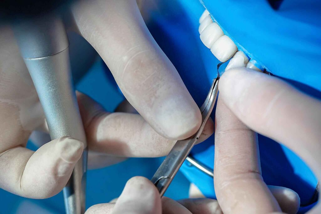
저는 간단한 계층 구조를 사용합니다.
위치가 우선이다. 적재는 그 다음이다. 이동은 세 번째다.
접는 종이 또는 호일을 사용하면 접촉 위치를 파악할 수 있지만, 가장 큰 잉크 자국을 보고 그것이 가장 큰 힘을 나타낸다고 무조건 단정 짓지는 않겠습니다.
미국 국립의학도서관(NLM)에 등재된 2012년 연구에 따르면, 아티큘레이팅 페이퍼의 표시 크기는 교합력의 백분율을 나타내는 신뢰할 수 없는 지표인 것으로 나타났다. 이전의 한 연구에서도 유사한 결론을 내렸는데, 표시 면적만으로는 실제 하중을 직접적으로 측정하는 지표로 간주해서는 안 된다는 것이었다.
그로 인해 그 물린 자국을 해석하는 방식이 달라집니다.
자국은 접촉의 증거입니다.
이것은 로드셀이 아닙니다.
환자의 정상적인 교합 상태부터 시작하십시오.
환자에게 즉시 과도한 움직임을 하도록 지시하지 마십시오. 먼저 환자가 자연스럽게 최대 교합 위치까지 입을 다물었을 때 어떤 현상이 발생하는지 확인하십시오.
제가 찾고 있는 것은:
목표는 모든 치아에 똑같은 흔적을 남기는 것이 아닙니다.
그건 마치 색칠공부 책 같은 치과 치료예요.
목적은 최종 수복 형태가 기능적 관계를 수정해야 할 정도로 변화시켰는지 여부를 파악하는 데 있습니다.
다음으로, 환자를 앞으로 데려오십시오.
6개, 8개 또는 10개 치아로 구성된 전치부 치료 사례는 단순히 절단면의 길이, 경사도, 두께 또는 안면-구개 방향의 위치가 달라졌다는 이유만으로도 전방 이동 유도 방향에 변화를 줄 수 있습니다.
또한, 하악 전치가 구개면이나 절단부와 맞물리는 위치가 바뀌게 된다면, 단 1밀리미터의 심미적 개선조차도 기능적인 문제를 초래할 수 있습니다.
전방 치아 접촉이 다음과 같은지 확인해 보고 싶습니다:
하지만 전방 접촉면을 무조건 무작정 갈아내지는 않겠습니다.
베니어는 수복물이지, 박물관 전시품이 아닙니다.
만약 수복 설계에 전방 유도 원리가 의도적으로 반영되었다면, 세라믹 치아 간 접촉이 시술자에게 불안감을 준다는 이유로 이를 제거할 경우, 의도적으로 계획된 기능을 훼손할 수 있다.
이제 옆으로 움직이세요.
개방교합, 군집 기능, 과거의 마모 양상, 치아 위치, 골격적 관계, 기존 후방 수복물, 그리고 비정상적인 구강 기능 등이 모두 적절한 치료 방법을 결정하는 데 영향을 미칩니다.
모든 베니어가 반드시 측면 접촉이 전혀 없어야 한다는 보편적인 규칙은 없습니다.
또한, 새로 접착된 개 치아 베니어가 작업면에서 발생하는 어떤 강한 접촉이든 무조건 견뎌내야 한다는 타당한 근거가 있는 규칙도 없습니다.
상황에 따라 달라집니다.
저에게 있어 경고 신호는 설명 없이 변경.
치료 전 환자의 치아 배열이 안정적이었던 경우, 새로 장착한 베니어가 설계나 예상에 없던 국소적인 교합 간섭이나 비교합 간섭을 유발한다면, 환자가 진료실을 떠나기 전에 이를 반드시 확인해야 합니다.
일부 환자들은 교과서적인 진찰 과정만으로는 관련 문제를 전혀 드러내지 않는 경우가 있습니다.
평소처럼 닫아 달라고 부탁하세요.
그들에게 자연스럽게 앞으로 몸을 기울여 달라고 부탁하세요.
필요한 경우, 말하기와 관련된 하악 위치 및 습관적인 하악 위치를 관찰하십시오.
가장자리 대 가장자리 관계, 심한 심교합, 현저한 전치부 마모, 또는 비정상적 구강 기능 사용 이력이 있는 환자의 경우, 단일 정적 교합 기록보다 기능적 범위(functional envelope)에 더 많은 주의를 기울여야 합니다.
바로 이 지점에서 다중 베니어 교합은 도식적인 접근 방식이 아닌 환자 맞춤형 접근 방식으로 전환됩니다.
| 시멘트 주입 후 확인 사항 | 왜 내 관심을 끄는가 | 연마하기 전에 확인해야 할 사항 |
|---|---|---|
| 교합 시 한 개의 베니어가 인접 치아보다 앞서 위치한다 | 조기 접촉 또는 장착 관련 변화의 가능성 | 폐쇄를 반복하고, 치아의 착석 상태, 접촉 부위 및 대합치를 확인하십시오. |
| 한 개의 중심치가 전방 이동에 주도적인 역할을 한다 | 집중적인 지도가 의도하지 않은 결과를 초래할 수 있다 | 승인된 설계 및 반대측 중앙부와 비교 |
| 새로운 비작동 간섭 | 하악의 움직임을 변화시키거나 하중이 집중될 수 있음 | 조정하기 전에 동작을 여러 번 반복해 보세요. |
| 무거워 보이는 접이식 종이 자국 | 마크의 크기만으로는 힘을 신뢰성 있게 정량화할 수 없다 | 위치, 반복적인 폐쇄, 촉각적 평가, 환자 피드백 및 필요한 경우 디지털 분석을 종합적으로 고려하십시오. |
| 접촉부는 최근 조정된 세라믹 위에 위치해 있습니다 | Surface의 원래 마감 처리가 손상되었을 수 있습니다 | 재질별 전용 시스템을 사용하여 점검하고 재광택 처리하십시오 |
| 환자는 한쪽 베니어에서 물었을 때 “가장 먼저 닿는다”고 말합니다 | 주관적인 정보는 실제 변화를 드러낼 수 있다 | 불만을 일축하기보다는 습관적인 폐쇄 여부를 다시 확인해 보십시오. |
| 시멘트 접착 후 여러 개의 전방 접촉부가 변경됨 | 단일 장치의 문제가 아니라 시스템 차원의 문제일 수 있다 | 한 개의 치아를 다루기 전에 완전히 복원된 구간 전체를 다시 점검하십시오. |
| 이갈이 증상 및 고밀도 세라믹 접촉 | 시간이 지남에 따라 기능적 위험이 높아질 수 있다 | 교합 계획 및 보호 장치 전략을 평가한다 |
그 마지막 행은 평소보다 더 많은 관심을 받을 만합니다.
베니어 업계에서는 세라믹의 강도에 대해 자주 언급하곤 합니다.
환자들은 굽힘 강도 수치를 저해하지 않습니다.
이것들은 수복물을 파손시킵니다.
2020년에 실시된 전향적 임상 연구에 따르면 64명의 환자에게 364개의 장석 베니어 시술을 시행했다, 이 중 이갈이 환자 40명을 포함하여, 최대 8년 동안 추적 관찰했다. 보고된 전체 생존율은 3년 차 93.7%, 5년 차 91%, 8년 차 87.1%. 이갈이 하위군에서, 교합 스플린트를 사용한 환자들은 사용하지 않은 환자들보다 생존율이 현저히 높았으며, 보고된 장기 생존율은 89.1% 대 63.9%였다.
그렇다고 해서 모든 이갈이 환자가 무조건 똑같은 교정 장치를 착용해야 한다는 뜻은 아닙니다.
이는 즉, 비정상적 기능(parafunction)을 부차적인 문제로 취급해서는 안 된다는 뜻입니다.
이전 임상 연구에서는 다음을 조사했다. 70명의 환자를 대상으로 한 323개의 도자기 라미네이트 베니어, 이갈이 증상이 있는 환자들에게 170개의 베니어를 시술했다. 연구진은 13건의 파절과 29건의 박리 사례를 기록했으며, 이 중 이갈이 증상이 있는 환자들에서는 8건의 파절과 22건의 박리 사례가 발생했다.
이러한 수치들이 베니어 시술을 피해야 한다는 근거가 되는 것은 아닙니다.
이는 기능을 존중해야 한다는 주장의 근거가 됩니다.
게다가 이런 상황들 때문에 “한 입 정도는 괜찮겠지”라는 식의 태도를 옹호하기가 점점 더 어려워지고 있다.
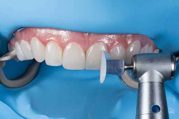
이 글에서 제가 가장 확고하게 생각하는 의견은 다음과 같습니다:
잉크를 쫓지 마십시오.
특정 접점이 원치 않는 간섭으로 반복적으로 확인될 경우, 조정이 필요할 수 있습니다. 하지만 세라믹을 제거해야 하는 이유는 기능적 문제가 확인되었기 때문이지, 단순히 한 개의 표시가 바로 옆의 표시보다 더 어둡게 보인다는 이유 때문은 아닙니다.
제 접근 방식은 보수적일 것입니다:
그 일곱 번째 단계에서 사람들이 걸려들곤 합니다.
간섭 요인 하나를 제거하면 힘의 분포가 달라진다.
물린 부위를 다시 한 번 확인해야 합니다.
세라믹 베니어의 교합 조정은 세라믹 표면을 변화시킵니다.
그게 중요합니다.
2014년에 실시된 한 실험실 연구에서는 조정 처리된 지르코니아 및 리튬 디실리케이트 시편과, 조정 및 연마 처리된 시편, 조정 및 유약 처리된 시편을 비교 분석했다. 임상적으로 의미 있는 결과 중 하나는 다음과 같았다. 조정된 리튬 디실리케이트와 지르코니아를 연마한 후, 대향 치아 법랑질의 마모가 감소했다..
이 연구는 체외 연구였을 뿐, 임상적인 베니어 수명 시험은 아니었으므로, 이 연구가 특정 환자의 예후를 입증한다고 단정 짓지는 않겠습니다.
하지만 이는 매우 실용적인 원칙을 뒷받침합니다:
세라믹을 연마할 경우, 표면을 적절히 마감해야 합니다.
눈에 띄게 교정된 부위를 그대로 두고 시술을 완료했다고 판단하는 것은 올바른 마무리 절차가 아닙니다.
Artist Dental Lab의 다음 주제에 관한 기사 광택 처리된 베니어와 유약 처리된 베니어의 비교 또 다른 중요한 점을 지적하고 있습니다. 표면 마감은 거칠기뿐만 아니라 수복물이 빛을 반사하는 방식에도 영향을 미칩니다. 따라서 과도한 수정은 기능적 문제뿐만 아니라 심미적 문제로도 이어질 수 있습니다.
일반적인 “도자기 연마” 지침은 현대 수복 치과 치료에 있어서는 너무 모호합니다.
이실리케이트 리튬, 장석질 도자기, 지르코니아는 서로 동일한 미세구조를 가지고 있지 않습니다.
따라서 보정 후 처리는 해당 세라믹 시스템에 대한 현행 지침에 따라 진행되어야 합니다.
IPS e.max는 단연코 최고의 기준입니다.
Ivoclar currently describes IPS e.max CAD as a lithium-disilicate glass ceramic and reports a mean biaxial flexural strength of 530 MPa 파단 인성이 2.11 MPa·m¹ᐟ². The manufacturer also documents both polishing and glazing routes, which is useful evidence against the simplistic idea that every adjusted E.max surface must be treated in one universal way.
For multi-unit lithium-disilicate cases, Artist Dental Lab’s comparison of monolithic versus layered E.max veneers is particularly relevant because adjustment does not occur in a vacuum. A monolithic incisal zone and a delicately layered incisal zone may not tolerate casual reshaping in the same esthetic way.
Feldspathic veneers can deliver exceptional optical detail, but thin ceramic plus delicate surface morphology leaves little room for thoughtless chairside reshaping.
If the contact problem requires substantial alteration of incisal length, palatal contour, or facial geometry, I would stop treating the issue as “just an occlusal adjustment.”
At some point, you are redesigning the restoration.
That may be better handled through laboratory correction or remake rather than progressively thinning ceramic until the bite mark disappears.
Zirconia follows another finishing logic.
Its oxide ceramic structure differs from silica-based glass ceramics, so finishing systems, polishers, and bonding protocols should not simply be borrowed from an E.max workflow.
Same appointment.
다른 화학.
Ceramic veneers can perform remarkably well when indication, enamel preservation, fabrication, bonding, and functional management are controlled.
A 2019 retrospective analysis followed 364 pressable lithium-disilicate laminate veneers in 41 patients for 10 years. The reported survival rate was 97.4%, with complications in 1.64% of restorations: fractures accounted for 0.55% and debonding for 1.09%.
Another long-term study of feldspathic porcelain veneers reported estimated survival around 96% at 10 and 20 years in its random sample when the veneers were bonded to prepared enamel.
That is excellent performance.
But I think survival statistics are often used too lazily.
A 97% survival figure does not mean clinicians can ignore one newly created contact because “E.max is strong.”
It means good outcomes are possible when the whole restorative system behaves well.
And occlusion is part of that system.
I would want the final record to answer several questions.
A functional check means very little if a restoration is not seated where it was intended to sit.
Margins, contacts, cement cleanup, and visual seating come first.
The patient should be able to close repeatedly without searching for a comfortable position.
A complaint such as “this tooth touches first” deserves investigation.
Do not immediately tell the patient they need to “get used to it.”
Sometimes adaptation is real.
Sometimes the restoration is high.
If the new incisal length changed anterior guidance, confirm that the change belongs to the treatment plan.
Look for new interferences rather than blindly pursuing a textbook occlusal pattern.
After ceramic adjustment and repolishing, inspect the restoration under more than one lighting direction.
A polished flat spot on one central incisor can alter the reflection pattern even if the shade remains unchanged.
Patients receiving several veneers need a maintenance strategy.
아티스트 덴탈 랩의 가이드 long-term veneer gloss, texture, and shade stability makes the useful distinction between a restoration merely remaining bonded and a restoration continuing to look and function well.
I agree with that distinction.
Survival is the floor.
Not the ceiling.
For a four-to-ten-unit anterior case, this is the sequence I would build into the clinical workflow:
Step 1: Confirm complete seating and cement removal.
Do not diagnose occlusion around residual resin or uncertain seating.
Step 2: Record habitual maximum intercuspation.
Use an appropriate contact indicator and repeat the closure enough times to distinguish a reproducible contact from noise.
Step 3: Compare anterior and posterior contact distribution.
Look for something that changed after cementation.
Step 4: Evaluate protrusive movement.
Identify the working path across the new incisal edges and palatal surfaces.
Step 5: Evaluate right and left lateral excursions.
Check intended guidance and investigate newly created interferences.
Step 6: Reproduce suspicious contacts before touching ceramic.
One blue dot is not a diagnosis.
Step 7: Perform the minimum justified veneer occlusal adjustment.
The objective is correction, not reshaping.
Step 8: Recheck the complete occlusion.
Every adjustment can redistribute contacts.
Step 9: Restore surface smoothness and gloss with the ceramic-specific finishing protocol.
Do not send roughened lithium disilicate, feldspathic porcelain, or zirconia back into function.
Step 10: Assess parafunctional risk.
For patients with bruxism, clenching, severe wear, or previous ceramic fracture, decide whether a protective appliance and closer follow-up belong in the plan.
Step 11: Document the final condition.
Clinical photographs, bite records when appropriate, notes about adjustment areas, material identification, and protection strategy make future troubleshooting far easier.
That is the difference between “checking the bite” and managing a restorative system.
A post-cementation occlusal check is the systematic verification of static and dynamic tooth contacts after veneers have been definitively bonded, using the patient’s actual seated restorations to identify premature contacts, guidance interferences, asymmetric loading, or newly created contact patterns that were not evident during the pre-bonding try-in.
It should include maximum intercuspation and clinically relevant excursive movements rather than one quick closure on articulating paper.
To check occlusion after veneer cementation, verify maximum intercuspation first, then evaluate protrusive, right and left lateral movements, edge-to-edge positions when relevant, and the patient’s habitual closure, comparing the new contact pattern with the planned anterior guidance and paying particular attention to isolated heavy or newly introduced contacts on ceramic.
Repeated verification matters because articulating-paper mark size alone should not be interpreted as a direct measurement of force.
Porcelain veneer occlusal adjustment is a conservative, material-specific process in which only confirmed interfering ceramic is reduced, the patient is repeatedly rechecked in static and excursive movements, and every adjusted surface is restored to an appropriate smooth polish or finish according to the ceramic manufacturer’s current instructions rather than left visibly or microscopically rough.
When the correction would significantly alter ceramic thickness, anatomy, or esthetics, laboratory correction or remake may be more defensible than aggressive chairside grinding.
Veneers may participate in protrusive or lateral guidance when that contact pattern was intentionally designed for the patient, but they should not be assigned or stripped of functional contacts by a universal rule; the clinician must judge contact location, force distribution, ceramic design, opposing dentition, parafunction, and the pre-treatment occlusal scheme together.
The target is controlled function, not automatically zero contact.
An occlusal splint is a protective removable appliance that may be indicated after veneer treatment for patients with diagnosed or suspected bruxism, especially when the restorative plan carries higher functional risk, because long-term clinical studies associate unmanaged parafunction with more veneer complications and show better survival in bruxism cohorts who consistently used protective splints.
A 2020 eight-year prospective veneer investigation found a marked survival difference between bruxism patients who used an occlusal splint and those who did not.
The best occlusal protocol for multi-veneer cases is a staged post-bonding verification that checks complete seating and cement cleanup, maximum intercuspation, protrusive and lateral guidance, edge-to-edge relationships when relevant, contact symmetry across the restored segment, patient comfort, and the final surface quality of any adjusted ceramic before the case is considered finished.
High-risk functional findings should then be incorporated into follow-up and protective-appliance planning rather than treated as isolated chairside observations.
A multi-veneer case should not be declared finished because the patient likes the mirror.
Finish the function.
Verify maximum intercuspation. Test protrusive and lateral movements. Investigate contacts that changed. Make ceramic veneer bite adjustment only when the interference is repeatable and clinically justified. And if ceramic is adjusted, restore its surface correctly.
The evidence gives us little excuse for casual work. Ten-year lithium-disilicate veneer survival can exceed 97% in well-controlled clinical series, yet bruxism data also show how dramatically functional risk can change outcomes.
That is the real lesson.
Strong ceramic helps.
Controlled occlusion helps more than marketing departments like to admit.
For clinics planning complex four-, six-, eight-, or ten-unit veneer cases, send your digital scans, opposing arch, bite records, stump shades, clinical photographs, approved smile design, material preference, and functional requirements to 아티스트 치과 실험실.
The laboratory can build the ceramic.
But the final post-cementation occlusal check decides how that ceramic enters the patient’s real mouth.