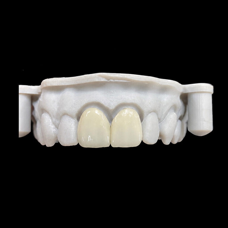
Veneer Preparation for Protrusive Teeth: Managing Profile and Lip Support
Profile comes first.
When maxillary incisors already sit facially, adding even a thin ceramic layer can increase apparent tooth projection, push the lip forward, thicken the cervical contour, and turn a technically accurate veneer case into an obvious facial mismatch.
Why would adding material automatically solve a volume problem?
It does not.
That is the hard truth behind porcelain veneer preparation for protrusive teeth. Veneers can alter visible crown form, close selected spaces, soften rotations, change line angles, and visually reduce apparent width. But they do not move roots. They do not correct a skeletal discrepancy. And they cannot promise lip retraction when the underlying incisors remain in the same position.
I have a strong opinion about this: a veneer case should not be called conservative merely because the preparation depth is small. A no-prep restoration that makes an already prominent tooth more prominent may conserve enamel while damaging the facial result.
Real conservation requires control of both biology and volume.
Protrusive Teeth Are a Three-Dimensional Problem
“Protrusive teeth” can describe several very different conditions:
A normally positioned root with a facially prominent crown
Mild incisor proclination
Severe dentoalveolar proclination
Bimaxillary protrusion
Crowding that rotates part of the crown facially
Spacing combined with labial inclination
A thick or overcontoured existing restoration
A skeletal pattern creating a convex profile
Lip incompetence related to tooth position, skeletal form, or soft-tissue behavior
These conditions should not receive the same veneer preparation.
A patient may present with straight-looking teeth in a frontal photograph yet show excessive projection in profile. Another may have obvious proclination but a thick, well-supported lip that disguises it. A third may have only one facially displaced lateral incisor.
The photograph lies easily.
Profile views, three-quarter views, intraoral scans, cephalometric information when indicated, lip posture at rest, natural speech, maximum smile, overjet, root position, and the existing enamel envelope all change the diagnosis.
A 2025 three-dimensional experimental study used temporary acrylic additions to simulate incisor protrusion in ten young adults. The researchers found an almost linear relationship between added thickness and perioral soft-tissue movement. Isolated upper-incisor protrusion displaced an upper-lip landmark by approximately 0.65 mm for every 1 mm of simulated advancement, while combined upper- and lower-incisor protrusion produced lower-lip displacement of approximately 0.76 mm per millimeter. Individual response still varied with lip thickness and anatomy.
That does not give clinicians a universal conversion formula. It proves something more useful: small changes in facial dental volume can produce measurable lip changes.
Ceramic thickness matters.
Can Veneers Correct Protrusive or Proclined Teeth?
Veneers can correct the appearance of selected mildly protrusive teeth when the planned restoration remains inside an acceptable facial envelope. They are much less convincing when the patient expects root movement, meaningful lip retraction, correction of severe overjet, or elimination of a convex profile.
This distinction is often blurred during cosmetic consultations.
What Veneers Can Change
A carefully planned veneer may alter:
Visible crown inclination
Facial convexity
Transitional line angles
Apparent tooth width
Incisal-edge position
Tooth length
Contact position
Embrasure progression
Surface texture and light reflection
Limited spacing or rotation
The visual relationship between central and lateral incisors
The laboratory can make a wide tooth appear narrower by moving the reflective line angles inward. That approach is explained in Artist Dental Lab’s guide to transitional line angles and optical contouring. It changes what the eye reads without demanding an equally large physical reduction in tooth width.
What Veneers Cannot Change
A veneer cannot predictably:
Retract an incisor root
Reduce a skeletal maxillary prominence
Correct severe bimaxillary protrusion
Eliminate lip incompetence caused by tooth or jaw position
Create orthodontic space
Correct an unstable overjet
Move the entire dental arch posteriorly
Guarantee a flatter profile
This matters because veneers for proclined teeth are sometimes sold as “instant orthodontics.” I dislike that term. It suggests biological movement where only restorative camouflage is occurring.
In selected cases, camouflage works. A 2025 case report documented lithium-disilicate veneers for a 37-year-old woman with anterior spacing and proclination. The reported outcome was favorable after conservative preparation and structured planning. But it was one patient, not permission to treat every protrusive case with ceramic.
Case reports show possibility.
They do not establish predictability.
Veneers, Orthodontics, or Combined Treatment?
The right question is not, “Can a veneer fit on this tooth?”
Almost anything can be made to fit after enough reduction.
The better question is, “Can the proposed final contour improve the smile and profile while preserving a defensible amount of enamel?”
Clinical condition
Primary problem
Veneer-only outlook
More defensible route
Mild facial crown prominence with favorable root position
Crown form and reflection
Potentially suitable after mock-up testing
Conservative mock-up-guided veneers
Mild spacing with limited proclination and stable occlusion
Space distribution and crown shape
Possible in selected cases
Veneers after facial and functional verification
Moderate or severe incisor proclination
Tooth and root position
High overcontour or reduction risk
Orthodontics before restorative treatment
Bimaxillary protrusion with lip incompetence
Both arches and soft-tissue profile
Poor veneer-only indication
Orthodontic or surgical-orthodontic assessment
One facially displaced anterior tooth
Local position discrepancy
Case-dependent
Limited orthodontics, restorative treatment, or both
Dark substrate on an already facial tooth
Masking plus limited space
High preparation and bulk risk
Position correction, material reassessment, or broader restorative plan
Deep bite, edge-to-edge contact, or heavy parafunction
Functional loading
Elevated fracture and debonding risk
Occlusal or orthodontic correction before veneers
Patient demands visible lip retraction
Soft-tissue profile
Veneers cannot promise the requested result
Orthodontic evaluation and documented consent
Orthodontic treatment changes the foundation. Veneers change the shell.
A prospective orthodontic study of adults with Class I bimaxillary protrusion reported incisor and lip retraction, an increased nasolabial angle, and reduced circumoral convexity after treatment. Those are profile changes produced by moving teeth, not by reshaping their facial surfaces.
So when a patient’s priority is lip position rather than tooth color or shape, an orthodontic opinion is not an inconvenience. It is part of honest diagnosis.
Porcelain Veneer Preparation Must Begin With the Final Contour
Traditional preparation begins with the tooth.
Defensible preparation begins with the approved result.
For protrusive teeth, I would not recommend placing generic depth-cutting burs against the untreated facial surface and assuming that a standard reduction will produce sufficient space. The existing surface may already be too far facially. Following it simply reproduces the problem at a slightly smaller scale.
The sequence should be reversed:
Establish the acceptable facial and incisal position.
Create a diagnostic wax-up or digital design.
Transfer it through a physical mock-up.
Evaluate the patient’s profile, lip posture, speech, smile and occlusion.
Adjust the proposed contour.
Prepare through the approved mock-up.
Verify remaining enamel and restorative space.
Rescan the preparation and approved design separately.
This is why the site’s framework for five measurable veneer design goals starts with facial integration, tooth proportion, gingival architecture, optical identity, and function rather than ceramic selection.
Reduce the Tooth That Exists, Not the Tooth You Wish Existed
The facial reduction may need to vary by region.
A tooth with cervical prominence may require more controlled reduction in the cervical or middle third. An incisal third that already projects excessively may need a different path of draw and facial transition. A rotated tooth may require asymmetric preparation.
Uniform preparation is not always conservative preparation.
But aggressive reduction creates its own problem: dentin exposure.
A systematic review and meta-analysis published in 2024 reported approximately 99% survival and 99% success, with reported ranges of 98% to 100%, for veneers bonded to enamel in the included clinical studies. Outcomes were less favorable when the bonding substrate shifted away from enamel.
The number should not be used as a sales promise. Study populations, materials, follow-up periods, operator skill, occlusion, and failure definitions differ.
Still, the direction is difficult to ignore.
Enamel is valuable.
No-Prep Is Not Automatically the Best Veneer Design
No-prep veneers attract patients because they sound reversible, painless, and biologically gentle. In favorable cases, minimal-prep and no-prep approaches can perform well.
But prominent teeth are usually not favorable volume cases.
A prospective study followed 186 conventional and no-prep or minimally invasive veneers in 35 patients for a mean of nine years. Importantly, the investigators excluded patients with inadequate hard tissue, severe discoloration, and other conditions that could compromise isolation or bonding.
That selection detail matters.
Good results from selected minimal-prep cases do not prove that ceramic should be added to a tooth whose profile is already excessive. Artist Dental Lab’s discussion of ultra-thin veneer limitations makes the same practical point: reducing ceramic thickness reduces the room available for masking, contour correction, and technical adjustment.
Thin is a dimension.
Conservative is an outcome.
Lip Support Must Be Tested in Motion
Lip support in cosmetic dentistry is often discussed as though it were a simple before-and-after measurement.
It is not.
Lip posture is influenced by:
Incisor position
Veneer facial thickness
Lip thickness
Muscle tone
Skeletal relationship
Tooth display at rest
Vertical dimension
Incisal-edge length
Arch form
Upper- and lower-incisor relationships
Habitual lip strain
Speech and smiling dynamics
A patient may like a fuller smile during a posed photograph yet dislike the same contour at rest. Another may accept additional support in the central-incisor region but object to cervical fullness near the lateral incisors and canines.
That is why the provisional or mock-up must be assessed from several views.
The Profile Test
Record the patient:
At rest
With relaxed lip closure
During a natural smile
During a maximum smile
From the right and left profiles
From both three-quarter views
While speaking
During “F,” “V,” “S,” and “M” sounds
Look for increased lip strain, loss of natural closure, a more convex profile, excessive upper-lip projection, or a shelf-like cervical transition.
A retracted intraoral image cannot reveal these failures.
The Thickness Test
Do not evaluate only whether the mock-up looks “good.” Compare the intended facial contour with the existing tooth using the preoperative scan, wax-up, mock-up scan, and planned preparation.
Ask measurable questions:
How much volume is being added at the cervical third?
Is the middle-third convexity moving facially?
Is the incisal edge being lengthened or moved forward?
Can the desired ceramic thickness be achieved while remaining mostly in enamel?
Is masking required?
Does the proposed contour increase or reduce lip support?
Where does the restoration first contact the lip?
I would also test more than one design when profile risk is present. A difference of 0.3 or 0.5 mm may seem trivial on a CAD screen. It may not feel trivial to the patient’s lip.
Ceramic Selection Cannot Rescue a Poor Spatial Plan
Material selection should follow the preparation and optical problem.
It should not be used to avoid them.
Lithium disilicate, commonly represented chemically as Li₂Si₂O₅, provides a useful balance of translucency, strength, CAD/CAM compatibility, and controlled thickness for many anterior cases. Artist Dental Lab’s E.max veneer workflow lists conservative anterior restorations, shape correction, and moderate color improvement as case-dependent uses, with material selection tied to preparation design, shade, stump shade, margins, and occlusion.
Feldspathic porcelain can offer exceptional optical control and delicate surface characterization when substantial enamel remains and the substrate does not require heavy masking. Lithium disilicate may provide more mechanical reserve and workflow repeatability.
Neither material eliminates the spatial equation.
The site’s comparison of E.max and feldspathic veneers for ultra-thin cases correctly frames the choice around substrate color, ceramic thickness, occlusion, enamel availability, and the technician’s ability to manage light.
The Masking Trap
A protrusive tooth with a dark stump creates conflicting demands:
The restoration should not become more facially prominent.
The ceramic needs enough thickness or opacity to control the substrate.
The clinician wants to preserve enamel.
The patient wants a lighter shade.
The laboratory needs room for believable translucency and texture.
Something must give.
Reducing too little may produce excessive contour or inadequate masking. Reducing too much may expose dentin and weaken the bonding situation. Choosing a highly opaque ceramic may control color but produce a flat, high-value result.
This is not merely a material decision. It is a consent decision.
The patient should understand the trade-off before preparation.
Optical Contouring Can Reduce Apparent Prominence
Not every protrusive-looking tooth needs the same amount of physical correction.
Sometimes the eye is reacting to reflection.
A broad, flat facial surface creates a wide reflective zone. Strong gloss can make that surface appear even larger and brighter. Moving transitional line angles inward, softening the facial highlight, controlling surface texture, and adjusting embrasures may reduce apparent bulk without radically reducing physical width.
This is especially useful when enamel preservation limits how much tooth structure can be removed.
But optical contouring has limits.
It cannot hide a restoration that physically pushes the lip forward. It cannot correct a cervical emergence profile that resembles a ledge. And it cannot make an excessively facial incisal edge disappear in profile.
Tooth proportions also affect the result. Artist Dental Lab’s guide to veneer width-to-length ratios explains how ratios, line angles, gingival position, lips, and surface reflection work together. The article treats the commonly cited 78% to 82% range as a checkpoint rather than a compulsory design formula.
That is the correct attitude.
Numbers guide judgment. They do not replace it.
What the Laboratory Must Receive
A laboratory cannot manage facial profile from one cropped intraoral scan.
For veneer preparation involving protrusive teeth, the submission should include:
Full-face photograph at rest
Natural-smile and maximum-smile photographs
Right and left profile photographs
Three-quarter facial views
Retracted frontal and lateral views
Preoperative full-arch scan
Prepared full-arch scan
Opposing arch
Verified bite record
Approved wax-up, mock-up, or provisional scan
Natural-smile and speech video
Final shade
Individual stump shades
Preparation photographs
Existing and target incisal-edge positions
Overjet and overbite information
Protrusive and lateral contact notes
Bruxism or clenching history
Planned material
Areas where facial bulk must not increase
Written approval of any accepted compromise
The lab prescription should not say only “reduce protrusion.”
That instruction is vague and potentially dangerous.
A useful instruction might read:
Teeth #6–11: reproduce the approved mock-up while maintaining the preoperative upper-lip position. Do not increase cervical facial volume on #7–10. Move the apparent facial line angles inward on #8 and #9. Preserve the approved incisal-edge position. Use the profile photographs and mock-up scan as controlling references. Contact the clinic if material thickness cannot be achieved without increasing facial contour.
Patients should be told plainly that veneers correct visible crown form, not underlying tooth position.
For a protrusive case, informed discussion should cover:
The orthodontic alternative
Whether orthodontics could improve lip position
Expected enamel removal
Possible dentin exposure
The risk of increased facial bulk
The masking-versus-thickness conflict
The limits of restorative camouflage
Whether the mock-up changes lip closure
Functional risks
Material limitations
The possibility that the desired profile cannot be achieved with veneers alone
A static digital smile simulation should not be treated as proof.
It can show color, tooth length, and frontal alignment. It cannot reliably prove speech, lip strain, muscle adaptation, bonding substrate, ceramic thickness, or long-term functional behavior.
And this is where the industry often becomes uncomfortable. Patients are sold the image before the spatial problem has been solved.
That order should be reversed.
FAQs
Can veneers correct protrusive teeth?
Veneers can correct the visible shape of mildly protrusive or proclined teeth, but they cannot move roots, reduce skeletal protrusion, or reliably retract the lips; treatment is appropriate only when diagnostic records, occlusion, enamel availability, and an intraoral mock-up show that ceramic can improve appearance without creating excessive facial bulk.
Cases involving marked proclination, lip incompetence, severe overjet, or a convex profile usually deserve an orthodontic assessment before irreversible porcelain veneer preparation begins.
How should protrusive teeth be prepared for veneers?
Porcelain veneer preparation for protrusive teeth is a mock-up-guided reduction strategy that creates uniform ceramic space while preserving as much enamel as possible, especially in the facial and cervical thirds, and it must be planned from the intended final contour rather than from a generic depth-cut number.
The reduction may be asymmetrical when individual teeth are rotated or positioned facially. Preparation photographs and a reduction map should be sent to the laboratory.
Do veneers increase lip support?
Veneers can increase lip support because every added fraction of a millimeter changes the facial contour contacting the lips, although the response varies with tooth position, veneer thickness, lip thickness, muscle tone, and whether one or both arches are altered; therefore, static smile photographs alone are not enough.
The proposed contour should be tested at rest, in profile, during speech, and during natural and maximum smiling before the definitive ceramic is fabricated.
When is orthodontics better than veneers for protrusive teeth?
Orthodontics is generally preferable when protrusive teeth reflect root position, marked incisor proclination, lip incompetence, a convex profile, crowding, unstable overjet, or insufficient restorative space, because tooth movement can change the dental foundation while veneer preparation only changes the external crown contour.
Limited orthodontics may also reduce the amount of enamel removal required before final veneers, creating a more favorable restorative and bonding condition.
What is the best veneer design for protruding teeth?
The best veneer design for protruding teeth is the least bulky, enamel-preserving design that has been approved in a facially evaluated mock-up, provides sufficient ceramic thickness, maintains clean emergence profiles, and respects phonetics and anterior guidance; no-prep veneers are not automatically conservative when they add unwanted volume.
Material, opacity, facial reduction, line angles, surface texture, and incisal position must be coordinated rather than selected independently.
What records should be sent to the dental laboratory?
A dental laboratory needs full-arch preoperative and preparation scans, the opposing arch, a verified bite, profile and three-quarter photographs, natural-smile video, approved mock-up or provisional scan, final shade, stump shade, preparation map, incisal-edge targets, functional notes, and explicit instructions describing which contours may be reduced or preserved.
Without facial and approved-design records, the technician can evaluate fit and margins but cannot reliably determine whether the final veneers will support or overproject the lips.
Send the Profile, Not Just the Preparations
Before preparing a protrusive anterior case, stop and test the proposed result in the patient’s moving face.
Confirm the profile. Confirm relaxed lip closure. Confirm the cervical contour. Confirm speech. Confirm the available enamel. And confirm that the patient understands the difference between restorative camouflage and orthodontic correction.
Then send the laboratory a complete record.
For a technical file review, material discussion, trial case, or customized anterior veneer plan, submit the preoperative and preparation scans, verified bite, profile photographs, approved mock-up, stump shades, and written contour targets through the Artist Dental Lab contact page.