Interproximal Extension for Veneers: Hiding Margins Without Overcontouring
Margins change everything.
A veneer can be thin, beautifully layered, correctly shaded, and technically polished, yet if its proximal finish line sits directly in the observer’s line of sight or forces the technician to build an oversized contact, the restoration can announce itself before the patient even smiles fully.
So why do we still treat interproximal extension as a minor detail of veneer preparation?
I think part of the problem is that clinicians are often taught preparation depth in tenths of a millimeter while the laboratory is forced to solve the final restoration in three dimensions. The dentist sees a finish line. The technician sees ceramic thickness, contact position, path of insertion, reflective width, emergence profile, masking demand, and whether there is physically enough room to make the restoration look like a tooth rather than porcelain added onto one.
That distinction matters.
Interproximal veneer preparation should extend only as far as required to move the margin away from an esthetically vulnerable viewing zone, provide the intended proximal contour, and give the ceramic adequate space without sacrificing unnecessary enamel.
Not farther.
Not automatically through every contact.
And certainly not because a preparation diagram says so.
The Real Purpose of Interproximal Veneer Preparation
The proximal portion of a veneer preparation has several jobs at once.
It may need to:
Hide the ceramic-tooth junction from frontal and oblique viewing angles
Allow the laboratory to reposition or reshape the contact
Provide ceramic thickness for masking a discolored substrate
Permit correction of rotated or misaligned teeth
Support diastema closure
Preserve cleansable embrasures
Maintain a usable path of insertion
Keep as much bonding surface in enamel as the treatment objective allows
That is already more complicated than “break contact” versus “do not break contact.”
The hard truth?
A preparation can be conservative on paper and still produce an aggressive-looking restoration.
Artist Dental Lab’s discussion of enamel preservation in veneer preparation makes an important distinction: preserving enamel is valuable, but refusing to create the restorative space needed for correct contour can leave the technician choosing between excessive thickness, poor masking, compromised seating, or an overcontoured facial surface.
The target is therefore not the smallest preparation imaginable.
It is the smallest defensible preparation for the restoration being requested.
Hiding the Margin Is Not the Same as Extending the Margin
This sounds obvious.
It isn’t.
Imagine an anterior veneer whose mesial finish line ends just facial to the existing contact. From straight ahead, the margin may disappear reasonably well because the neighboring tooth blocks direct visibility.
Now rotate the tooth slightly. Add a dark proximal substrate. Move the planned contact. Close a diastema. Increase tooth width.
The geometry changes immediately.
A margin that was visually protected in one case can become exposed in another.
This is why proximal veneer extension should be planned relative to the final restoration, not merely the original tooth.
The line of sight test
I prefer thinking about margin position as a visibility problem rather than a fixed-distance problem.
Ask:
Can the margin be seen from the frontal smile view?
Can it be seen from a normal three-quarter angle?
Does the finish line sit inside the reflective facial zone?
Will the final ceramic require added bulk over that margin?
Does the planned contact move mesially, distally, facially, or lingually?
Those questions tell us more than an arbitrary number of millimeters.
There is no credible universal rule saying that every veneer margin must extend exactly 0.5 mm, 1.0 mm, or 2.0 mm past a contact.
Case geometry wins.
Three Levels of Proximal Veneer Extension
In practical terms, I separate proximal veneer preparation into three broad approaches.
Preparation Approach
Typical Situation
Main Advantage
Main Risk
Finish facial to contact
Minimal shape or shade correction with favorable tooth alignment
Maximum enamel conservation
Visible transition or limited contour control
Extend into contact region
Moderate shape correction or improved margin concealment
Better ceramic transition and contact control
Accidental contact opening or limited lab access
Extend lingual to contact
Diastema closure, major contact relocation, rotation correction, strong proximal discoloration
Maximum freedom for contour and hidden margin placement
More tooth reduction, dentin exposure, difficult hygiene if overbuilt
None of these deserves the title “best veneer preparation design” by itself.
The indication decides.
When stopping facial to the contact makes sense
If tooth position is favorable, the existing contact is acceptable, the substrate does not need heavy masking, and very little proximal contour change is required, extending farther simply to follow a routine may remove sound enamel without delivering an equivalent benefit.
Keep it simple.
But conservative preparation stops being conservative when the laboratory has to add excessive ceramic outside the original tooth envelope to achieve the requested shape.
That is where no-prep and minimal-prep rhetoric can become misleading.
When extending into or through the contact makes sense
Greater proximal extension becomes more rational when the restoration must substantially alter:
Mesiodistal width
Contact location
Tooth rotation
Diastema dimensions
Black-space appearance
Proximal masking
Symmetry between adjacent veneers
Breaking a contact can also give the laboratory more freedom to establish the final contact inside the ceramic rather than being forced to terminate porcelain beside an untouched proximal wall.
But more freedom is not automatically a better result.
It merely gives the technician more design territory.
The 2024 Margin Study Deserves Attention
One of the more relevant pieces of recent evidence comes from researchers associated with the University of Otago Faculty of Dentistry.
Bennani, Aarts, and Senthilkumar compared three laminate veneer preparation forms: feather edge, shoulder, and shoulder with wings. Their 2024 Journal of Prosthetic Dentistry study used CAD/CAM veneers and scanning electron microscopy to assess absolute margin discrepancy and marginal overhang.
The PubMed record for the 2024 preparation-design study reports that the shoulder-with-wings design produced the smallest cervical absolute margin discrepancy and overhang, while proximal measurements were comparable with the shoulder design.
This was an in-vitro investigation, so I would not turn it into a universal clinical recipe.
But the result matters.
Preparation geometry changes what the laboratory can manufacture.
That should not be controversial.
The Overcontouring Trap
Now we reach the part that is routinely underestimated.
Veneer overcontouring occurs when the final restoration projects farther facially or proximally than the intended natural tooth envelope because insufficient preparation space, incorrect margin placement, contact design, or ceramic buildup forces excess restorative volume.
It can happen with very thin veneers.
Thickness alone does not define overcontouring.
Position does.
A 0.5 mm veneer can still look bulky
Suppose a clinician preserves almost the entire facial surface because the case is marketed as “minimal prep.”
Good intention.
But the approved design requires the central incisor to move facially only slightly while its proximal surface must move significantly to close a space.
If no corresponding proximal reduction exists, ceramic must be added outward.
The contact broadens.
The transition line moves.
The deflective zone disappears.
Suddenly the veneer looks wider and flatter even though its measured thickness sounds impressively conservative.
That is optical overcontouring.
Artist Dental Lab’s guide to transitional line angles in veneers explains why: the mesiofacial and distofacial line angles define the central reflective zone. Move those lines outward and the tooth can appear wider; move them inward and the apparent facial width can become narrower without changing the actual mesiodistal dimension.
That is why I become skeptical whenever someone evaluates veneer preparation only with a reduction gauge.
Calipers cannot measure visual mass.
Where Veneer Overcontouring Usually Starts
Several patterns repeatedly create trouble.
1. The margin stays too facial
The technician must transition from ceramic to tooth while still inside a highly visible surface.
To hide that transition, porcelain gets feathered, blended, or thickened.
Now the restoration is being designed around the margin rather than around natural anatomy.
2. The contact cannot be moved
If the original contact remains intact but the treatment plan calls for substantial width correction, the technician has limited room to reposition the final interproximal contour.
The result can become a compromise between the old anatomy and the proposed smile design.
3. The preparation ignores the wax-up
This one bothers me most.
Why approve a new tooth position and then prepare according to the old tooth?
A preparation should be evaluated relative to the proposed restoration volume. Otherwise, “minimal preparation” becomes random preparation.
4. The lab receives no preparation photos
A digital scan can show geometry.
It does not always explain the clinical intention.
Artist Dental Lab’s anterior veneer case submission guide recommends providing the prepared arch, opposing arch, bite, preoperative scan, approved provisional or mock-up, preparation photographs, stump shade, final shade, facial photographs, and functional information rather than expecting the technician to reconstruct the plan from one STL file.
That is not paperwork.
That is design information.
Enamel Preservation Still Matters
We should not solve every esthetic problem by extending farther.
Ceramic veneers derive much of their predictability from adhesive bonding, and enamel remains an especially desirable bonding substrate.
A 2012 literature review from the University of Birmingham examined 24 publications and concluded that there was reasonable evidence that extending veneer preparation into dentin adversely affected survival. The authors explicitly stated that the ideal veneer preparation remains within enamel.
That creates the real tension in interproximal veneer preparation.
We want enough extension.
But not unnecessary extension.
So the correct question is not:
“Should I break the contact?”
It is:
“What is the minimum proximal extension that allows the planned restoration to be fabricated with acceptable visibility, contact position, ceramic thickness, contour, cleansability, and enamel preservation?”
That is a much harder question.
It is also a better one.
Long-Term Data Punishes Sloppy Margin Decisions
Long-term veneer performance is generally impressive, but the headline survival percentage hides clinically useful detail.
A 2021 systematic review evaluated 25 studies and 6,500 porcelain laminate veneers. The reported estimated cumulative 10-year survival rate was 95.5%. Fracture was the most common complication, followed by debonding. The review also discussed the greater bonding concern when preparations involve larger areas of dentin.
Good numbers.
But survival is not the same as perfection.
A restoration may survive while developing marginal staining, roughness, contour problems, repair needs, or other changes that never qualify as catastrophic failure.
That distinction becomes obvious in older clinical data.
What an Actual 10-Year Veneer Trial Found
Peumans and colleagues followed porcelain veneers placed on 87 maxillary anterior teeth in 25 patients.
At five years, an average of 92% of restorations remained clinically acceptable without intervention. At 10 years, that figure had fallen to 64%, although only 4% required replacement.
The more revealing numbers?
11% showed porcelain fractures and 20% developed large marginal defects.
That should change how we think about veneer margin placement.
A margin is not merely where the technician stops ceramic.
It is an interface expected to survive years of moisture, loading, hygiene, staining exposure, adhesive aging, and repeated professional maintenance.
Research Still Disagrees About “The Best” Veneer Preparation
Here is another reason I distrust universal preparation doctrines.
A 2017 systematic review and meta-analysis screened 415 publications and included 10 studies comparing veneer preparation designs. Based on time-to-event data, preparations with incisal coverage showed a higher failure risk than those without coverage, with a reported hazard ratio of 1.81.
Yet the 2021 long-term review of 6,500 veneers reported the opposite direction for incisal coverage.
Contradictory evidence does not make research useless.
It tells us something more useful: preparation shape cannot be divorced from material, bonding substrate, occlusion, operator technique, tooth condition, and case selection.
The same skepticism should be applied to proximal extension.
How Far Should Veneers Extend Interproximally?
There is no universal millimeter measurement.
That is my answer.
A veneer should extend interproximally far enough to satisfy the planned esthetic and restorative change while stopping as early as possible once the margin can be positioned predictably, the contact can be created correctly, sufficient ceramic space exists, and unnecessary dentin exposure can be avoided.
Several variables change the answer.
Existing tooth alignment
A well-aligned tooth requiring only color correction usually needs less proximal intervention than a rotated tooth being visually repositioned.
Diastema closure
Closing space changes tooth width and contact geometry.
The preparation may therefore need to move farther lingually so the restoration can wrap into the proximal surface rather than simply adding a slab of ceramic facially.
Substrate color
A dark preparation that remains visible beside a translucent proximal ceramic margin can expose the restoration-tooth transition.
Greater extension may be justified when the esthetic benefit outweighs additional reduction.
Existing proximal restorations
Composite restorations, cracks, caries, or irregular proximal anatomy alter where a predictable finish line can be established.
The 10-year Peumans study is a reminder that existing composite at veneer boundaries deserves attention rather than being treated as irrelevant background anatomy.
Contact relocation
If the technician needs to move the contact significantly, the preparation must provide enough freedom to build that new anatomy.
A ceramic contact cannot occupy space already occupied by tooth structure.
Physics remains stubborn.
Margin Placement Should Follow the Final Tooth, Not the Original Tooth
This is probably the single most useful principle in the entire discussion.
Start with the final contour.
Then determine where tooth structure conflicts with that contour.
Remove only what interferes.
This mock-up-driven mindset makes much more sense than preparing a standardized veneer shape and later asking the technician to force every case into it.
For clinicians working with lithium disilicate, Artist Dental Lab’s E.max veneer workflow also emphasizes case-dependent minimal-to-standard preparation, clear margin definition, reduction notes, shade information, and digital records rather than prescribing one reduction design for every anterior restoration.
And that is exactly how it should be.
Proximal Contour Is an Optical Problem Too
Dentists naturally focus on contact strength and floss resistance.
The laboratory sees another layer.
The contact has an optical consequence.
If the proximal facial surface stays too full, the deflective zone shrinks. More light returns toward the viewer. The veneer appears broader.
Then someone asks the technician to make it look narrower.
So gray stain gets added.
Or the line angle gets carved too aggressively.
Or the ceramic gets ground after delivery.
Wrong sequence.
Shape first. Color second.
If the restoration is physically overcontoured, pigment is camouflage.
Artist Dental Lab’s discussion of veneer incisal-edge adjustment and surface character demonstrates the related problem: grinding ceramic changes material volume, surface texture, and the geometry controlling reflected light at the same time.
The same principle applies proximally.
Correcting a bad contour after final ceramic finishing is harder than designing the contour correctly from the beginning.
Material Choice Changes the Amount of Freedom
Porcelain veneer preparation cannot be separated from the ceramic being prescribed.
Feldspathic porcelain
Feldspathic veneers can provide exceptionally delicate optical layering and thin ceramic transitions, making them attractive for highly conservative esthetic cases.
But thin ceramic is not magic.
If major masking, contact relocation, or substantial shape correction is needed, available restorative space becomes a serious design variable.
Lithium disilicate
Lithium disilicate, commonly represented by Li₂Si₂O₅, provides greater mechanical reserve than conventional feldspathic porcelain and supports pressed, milled, monolithic, cut-back, and layered workflows.
Still, material strength does not justify overcontouring.
A stronger bulky veneer is still bulky.
Zirconia
Zirconia may offer higher opacity and strength for selected cases, but optical behavior differs from glass ceramics.
Again, the preparation should follow the restorative objective.
Material does not rescue incorrect geometry.
What the Laboratory Needs Before Designing the Proximal Surface
If I were evaluating a difficult veneer case from the laboratory side, I would want six pieces of evidence before making major decisions about proximal anatomy:
Preoperative scan showing original tooth position and contacts
Prepared scan with readable proximal margins
Approved wax-up or provisional scan showing intended width and contact position
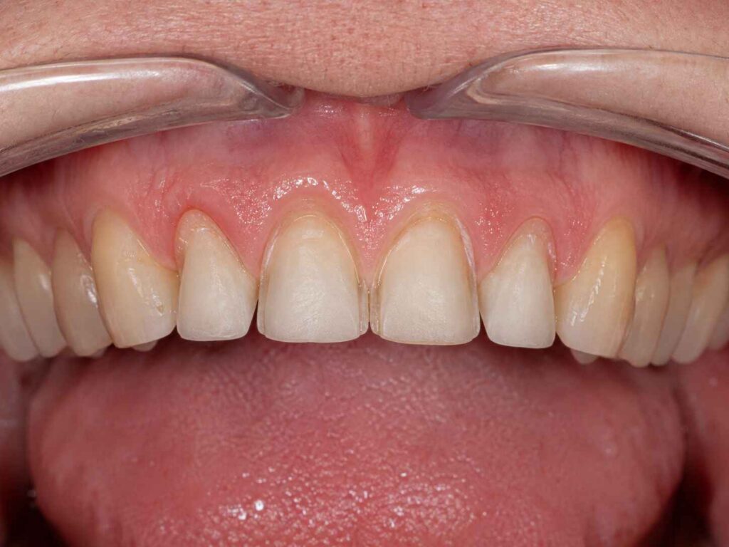
Preparation photographs showing enamel, dentin, discoloration, and margin visibility
Stump shade records for each prepared tooth
Frontal and three-quarter facial photographs showing how the proximal areas appear from normal viewing angles
Without these records, the technician may still produce a restoration.
But some of the design is now guesswork.
And “make the margins invisible” is not a laboratory prescription.
A Practical Decision Framework for Interproximal Veneer Preparation
Before extending farther proximally, I would ask these questions in order.
1. Is the existing contact staying where it is?
If yes, aggressive extension may provide little advantage.
If no, identify exactly where the final contact needs to move.
2. Is the veneer changing tooth width?
Minor width correction and major diastema closure are not the same treatment problem.
3. Will the existing margin be visible?
Evaluate frontal and oblique viewing angles.
Do not judge from the occlusal view alone.
4. Is there enough room for ceramic?
Compare the preparation to the approved wax-up.
If ceramic must be added outside the intended envelope simply because tooth structure occupies the necessary space, the preparation is under-reduced in the wrong location.
5. How much enamel remains?
Additional extension should earn its biological cost.
6. Can the final contour be cleaned?
A hidden margin surrounded by a bulky, plaque-retentive proximal contour is not a successful esthetic solution.
7. Can the laboratory clearly read the finish line?
Margins that disappear inside scan noise, irregular surfaces, or poorly captured subgingival anatomy are not magically improved because they are hidden from the patient.
The Best Veneer Preparation Design for Esthetics Is Case-Specific
I know that answer is less satisfying than a diagram.
But dentistry does not owe us simple answers.
A conservative facial preparation may be ideal for one central incisor and completely inadequate for the rotated lateral beside it.
A contact-preserving design can work beautifully when tooth position already supports the final contour.
A lingually extended preparation can make far more sense when closing a diastema, moving the contact, masking proximal discoloration, or changing tooth orientation.
The mistake is treating any one of these as doctrine.
Design backward.
Begin with the tooth the patient and clinician approved. Evaluate the space required to build it. Locate the margin where it can be finished, scanned, bonded, cleaned, and visually protected. Then preserve every additional micrometer of enamel that does not interfere with that goal.
That is genuine conservative veneer preparation.
Not simply doing less.
Doing only what the restoration actually requires.
FAQs
What is interproximal veneer preparation?
Interproximal veneer preparation is the controlled extension of a porcelain veneer preparation from the facial surface toward the mesial or distal surfaces so the clinician can position margins, modify contacts, manage tooth width, provide ceramic space, and hide restoration boundaries while preserving as much enamel as the planned esthetic correction reasonably allows.
It may stop facial to the contact, enter the contact region, or extend lingually beyond it depending on tooth position and the intended final restoration.
How far should veneers extend interproximally?
Veneers should extend interproximally only far enough to place the margin outside an undesirable visible zone, permit the planned contact and tooth contour, provide adequate restorative space, and create a finish line the clinician and laboratory can manage without removing additional healthy enamel that contributes no meaningful esthetic or restorative benefit.
There is no evidence-based universal millimeter distance appropriate for every veneer.
Should interproximal contacts always be broken for veneers?
Interproximal contacts should not automatically be broken for every veneer because contact opening is a case-dependent preparation choice used when additional laboratory access is needed for margin concealment, contact relocation, diastema closure, rotation correction, masking, or substantial contour change, rather than a mandatory feature of successful porcelain veneer preparation.
When the existing contact and tooth position already support the final design, preserving the contact may conserve more enamel.
How can veneer overcontouring be prevented?
Veneer overcontouring is best prevented by designing the desired final tooth first, comparing that contour with the existing enamel through a wax-up or mock-up, and selectively creating restorative space where ceramic would otherwise be forced outside the natural envelope, particularly at facial-proximal transitions, contact areas, cervical emergence zones, and transitional line angles.
The objective is controlled space, not indiscriminate reduction.
Where should veneer margins be placed interproximally?
Interproximal veneer margins should be positioned where they can remain visually protected, clearly recorded, accurately manufactured, predictably bonded, and accessible for long-term hygiene while allowing the technician to establish natural contact form and proximal curvature without creating excessive ceramic thickness or unnecessarily extending the finish line onto dentin.
The correct location therefore depends on the final tooth position rather than one standardized preparation diagram.
What is the best veneer preparation design for esthetics?
The best veneer preparation design for esthetics is the least invasive geometry that provides sufficient space for the selected ceramic, maintains favorable enamel bonding where possible, positions margins away from exposed viewing zones, gives the laboratory control of contacts and line angles, and reproduces the patient-approved tooth form without overcontoured facial or proximal surfaces.
Window, butt-joint, wrap, contact-preserving, and contact-breaking designs all have legitimate indications.
Can E.max compensate for insufficient veneer preparation space?
E.max cannot fully compensate for insufficient veneer preparation space because lithium disilicate may provide useful strength and optical flexibility, but the restoration still requires appropriate three-dimensional volume for contour, translucency, masking, contacts, margins, and emergence profile; placing ceramic where tooth structure already occupies the planned contour inevitably changes the restoration’s external form.
Material properties can expand design options.
They cannot repeal geometry.
What should be sent to the dental lab for an anterior veneer case?
An anterior veneer laboratory case should include the prepared and opposing arches, bite record, preoperative scan, approved provisional or wax-up, preparation photographs, stump shade and final shade records, facial and retracted photographs, restoration material, margin information, contact instructions, and functional notes so the technician can reproduce an approved plan instead of interpreting missing clinical information.
For difficult proximal extensions, preparation photos and an approved design scan are especially useful because they show both the available space and the intended final contact.
Plan the Margin Before the Ceramic
Interproximal extension should never become a contest to see how far a veneer can wrap around a tooth.
The better objective is narrower: hide what should not be visible, preserve what does not need to be removed, and create exactly enough space for natural ceramic anatomy.
If you are planning E.max, layered E.max, feldspathic, or multi-unit anterior veneers and want the laboratory to evaluate preparation margins, contact design, available restorative space, stump shade, and digital files before production, send the case records through the Artist Dental Lab contact page.
Send the preparation scan.
Send the approved design.
Send the photographs.
Then give the technician enough information to build the veneer inside the intended tooth envelope—not around an avoidable preparation compromise.