価格・サンプルに関するお問い合わせ
ジルコニアクラウン、二ケイ酸リチウムクラウン、ベニア、OEMサービス、あるいは修復物の卸売り注文を比較検討している歯科技工所、歯科医院、販売代理店、および調達担当者の方へ。.
取り扱い製品の種類、素材、月間販売数量、輸出先国、およびサンプルのご要望をお知らせください。営業チームが適切な対応策を講じることができます。.

プレッシャーはすべてを変える。.
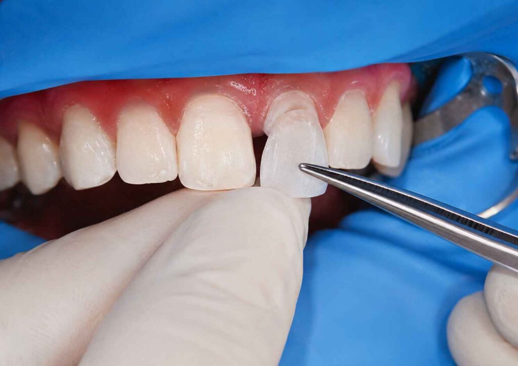
厚さ約0.3~0.5 mmの極薄ベニアを、その下にレジンセメントの層を敷いた歯に装着する際、施術者は単に「ベニアを所定の位置に押し込む」だけではありません。 圧力をかけることで、同時にレジンが移動し、余分な材料が押し出され、粘性が克服され、歯質形成の形状と相互作用し、修復物が意図した位置にどれだけ完全に到達するかが決まるのである。.
では、なぜベニアの接着手順では、これを依然として「指でしっかりと圧力をかける」という表現に簡略化しているのでしょうか?
その言葉が気になってしまう。.
“「Firm」は単位ではありません。.
どちらも「穏やか」とは言えない。“
また、修復体自体の厚さがわずか数百マイクロメートルに過ぎない場合でも、制御されていないセメント層が、修復システム全体の中で驚くほど大きな割合を占めることがあります。.
これは、以下の点においてさらに重要性を増す。 超薄型ベニア, 、ここでは、セラミックの厚さ、セメントの厚さ、基材の色、内部の適合性、および最終的な修復物の位置を、それぞれ独立した問題として扱うことはできない。.
2024年に行われた実験室での調査では、樹脂材料に対して標準化された 30 Nの荷重 ガラス板の間に形成された膜の厚さを測定した。試験対象となった21種類の修復用コンポジットおよび指定セメントについて、測定された膜厚はわずか 6 μm~200 μm.
座席定数は同じです。.
映画の作風がまったく異なる。.
この単一の結果だけでも、ベニアのセメント固定において最も安易な仮定の一つ――「同じ圧力をかければ、セメントの厚さは同じになる」という仮定――を覆すに十分だろう。.
いいえ、そうではありません。.
従来の修復方法では、わずかな誤差を吸収するための幾何学的な余裕がより多く確保されています。.
極薄のベニアではそうではありません。.
アーティスト・デンタル・ラボによる、 超薄型ジルコニアベニアの品質管理基準 おおよそ次のように説明しています 0.3~0.5 mm 修復物については、わずかな寸法変化でも修復物全体の相当な割合を占めることになるため、根本的に異なる検査上の問題となる。.
その表面の下でも、同じ理屈が当てはまります。.
顔面用セラミックの厚さが0.4 mmであると仮定する。.
つまり 400 μm.
さて、樹脂セメントの制御されていない部分が150~200 μmに達している状況を想像してみてください。.
セメントはもはや目に見えない技術的な細部ではありません。修復厚みにおいて重要な構成要素となっています。.
だからこそ、私は薄いベニヤ板を、単に次のように尋ねるだけで評価することなど決してないのです:
“「席は空いていましたか?」”
私はこう尋ねたいと思います:
それがベニアのセメント固定です。.
それ以外は、ただの陶器を押し売りしているに過ぎない。.
樹脂セメントには体積がある。.
充填剤の粒子が含まれています。.
粘性があります。.
稼働時間があります。.
そして、変位に耐える。.
ベニアが形成部に近づくと、余分なセメントは辺縁に向かって外側に流れ出さなければなりません。残りの隙間が狭くなるにつれて、材料が流れ出るスペースは少なくなります。.
抵抗が高まっている。.
内部の形状がきつい場合、近心側の接触が激しい場合、経時的に材料の粘度が上昇する場合、あるいは修復物が不均一に圧入された場合、見られる「着座抵抗」は、セラミック自体とはほとんど関係がない可能性があります。.
強く押し付けると、膜厚が薄くなる場合があります。.
しかし、時には強い圧力が、単に別の問題を覆い隠しているだけの場合もある。.
その違いは重要だ。.
「~と呼べる」と正当化できる普遍的な数は存在しない。 あらゆる超薄型ベニアに最適なセメント膜の厚さ.
研究によって、使用するセラミック、レジン系、歯面形成法、充填法、試料の形状、CADの設定、および測定手法が異なります。.
とはいえ、いくつかの知見は、あまりにも有用であるため、無視することはできない。.
| 証拠 | 実験の詳細 | 結果 | そこから得たもの |
|---|---|---|---|
| 2024年 樹脂素材フィルムに関する研究 | 21種類の材料;30 Nの標準荷重 | 膜厚の範囲は 6~200 μm | 着床力だけでは、最終的なセメントの厚さを予測することはできない |
| 2025年 CAD/CAM ジルコニアベニアに関する研究 | 60枚のベニア;フェザーエッジ、ショルダー、ショルダー・ウィズ・ウィングス | ショルダー形状は、フェザーエッジに比べて全体的なセメントの厚みが薄くなり、ウィング付きショルダーでは垂直層が最も薄かった。 | 調製時の形状によって、実際に生成されるセメント空間が変化する |
| 2023年の二ケイ酸リチウムベニアに関する研究 | 切歯24本;最小限の形成 vs ノープレップ | 頸部セメントの厚さは 87 ± 27 μm 最小限の加工を施したベニアと、 210 ± 89 μm 事前準備不要のベニア用 | “「ノープレップ」だからといって、必ずしも修復物の接合面が薄くなったり、より精密に制御されたりするというわけではない |
| 有限要素法を用いたベニア解析 | 以下に基づいてモデル化されたセメント層 10~180 μm | セメントの厚さが増すにつれて、モデル化された接着応力は増加した。著者らは、厚さがおよそ50 μm未満の層では、モデル化された接着破壊が軽減される可能性があると示唆した。 | 非常に厚いセメント層は、自動的に無害であると見なすべきではない |
2025年のオタゴ大学関連のジルコニアベニアに関する研究は、以下の点を測定したという点で特に興味深い。 60個のセメント固定式CAD/CAMジルコニアラミネートベニア, 、各調製群につき20例。ショルダー型およびショルダー・ウィング型の調製では、フェザーエッジ型に比べてセメントの全体的な厚みが薄かった。.
それは重要です。.
それは、臨床医が時折、指だけで解決しようとしてしまうある事柄を証明しているからです:
セメントの厚みは、セメント充填が始まる前から決まります。.
まずは準備の設計から始まります。.
2023年に行われたin vitro研究では、ヒトの上顎中切歯を対象に、最小限の歯質除去を施した二ケイ酸リチウムベニアと、歯質除去を行わない二ケイ酸リチウムベニアを比較した。.
低侵襲群では、 0.3 mmの切削.
セメント固定後、以下の期間を経て経時変化が生じます 6,000回の熱サイクル, そして 100 Nでの10万回の機械的サイクル, 、研究者らは頸部セメントの厚さを測定した。.
その数字は紛れもないものでした。.
最小限の処置で作成されるベニア: 87 ± 27 μm.
事前準備不要のベニア: 210 ± 89 μm.
平均的な頸部セメント厚の2倍以上。.
それは、準備を必要としないベニアがすべて悪いということを証明しているのでしょうか?
いいえ。.
これは、歯の保存と修復スペースが同じ概念ではないことを示している。.
ベニアはエナメル質をほとんど削らずに済む一方で、修復物が計画された輪郭を形成するのに十分なスペースが確保できないため、接合部の管理が難しくなることがあります。.
アーティスト・デンタル・ラボによる 隣接歯面へのベニアの延長および過形成 別の観点から同じ問題に言及している。すなわち、極端な保守主義は、意図的に確保された空間内ではなく、元の歯の輪郭の外側に修復用スペースを設けることを余儀なくされる可能性がある。.
準備を少なくすることが、必ずしもより良い歯科治療につながるわけではありません。.
正しいターゲットは 計画された復元作業に必要な、最低限の妥当な準備.
仕組みは単純そうに見える。.
圧力が高まると、より多くのセメントが外側に押し出される。.
セメント層が薄くなっていく。.
ベニアはさらに奥に配置されています。.
場合によっては、そうですね。.
しかし、実際の樹脂材料を取り入れると、これはもはや単純な話ではなくなります。.
2024年の研究では、同じ条件下で材料を比較し、 30 Nの実験荷重 以下の数値からフィルムの厚さを算出した 6 μm~200 μm.
一部の修復用コンポジットは、およそ 6~8 μm.
データセットの一部に含まれる流動性・注入性材料および指定セメントからは、およそ 15~27 μm 映画。.
その他の試験対象材料は、以下の範囲内に収まった。 81および200 μm.
それはとてつもない差ですね。.
そして、それは標準化された圧力にさらされた。.
だから、誰かが私にこう言うときは:
“「私たちは常に同じ着座圧をかけています。」”
私の回答は次のとおりです:
セメントの種類も知らなければ、それだけではほとんど何も分かりません。.
予熱した修復用コンポジットは、加熱によって操作性や粘度が変化するため、代替の接着材として注目を集めている。.
しかし、ここには落とし穴がある。.
見かけの粘度が低いからといって、あらゆる材料において予測可能な最終膜が得られるとは限りません。.
2024年の同調査では、初期粘度だけでは、熱改質セメンテーションの過程で最終的に形成される膜厚を確実に予測することはできないとの結論が導き出された。.
まさにその理由から、ブランドに依存しないセメンテーションに関するアドバイスは危険なものとなるのです。.
2つの材料は、加圧下ではどちらも流動性があるように見えるかもしれませんが、修復物が最終的な装着段階に近づくにつれて、それぞれ異なる挙動を示すことがあります。.
樹脂メーカーが検証済みの指示に従ってください。.
診療席で粘度測定のプロトコルを勝手に考案してはいけません。.
超薄型の中切歯用ベニアを、切縁の3分の1付近を1本の指で押さえる様子を想像してみてください。.
ベニアが微細な角度で傾くことがあります。.
セメントは、ある地域では別の地域よりも早く残ってしまう。.
一方のマージンが先に最終位置に到達する。.
反対側の領域には、より厚い樹脂層が残っています。.
すると、重合が始まります。.
こうして、セメントは座席の不均衡を記録してしまった。.
だからこそ、私はこう考えることを好むのです 制御された座面圧力, 、単に座面への圧力が高いというだけではない。.
修復にあたっては、以下の点に留意すべきである:
圧力は、そうした目的のために活用されるべきである。.
それを競争にしてはならない。.
指と歯の間に何があるか、忘れないように。.
2024年に実施された超高透光性多層ジルコニアベニアに関する調査では、破断抵抗性が表層の厚さによって異なることが明らかになった。報告された値には、およそ 742 N、1.00 mm そして 673 N、0.75 mm, 一方、0.50 mmの試験片は、その実験設計の下で、0.75 mmのグループと同等の破壊抵抗性を示した。.
役立つデータ。.
しかし、それをもってしても、セメント充填時の過度な圧力を正当化することはできない。.
破断試験荷重と臨床的な装着荷重は、まったく異なる疑問に答えるものです。.
これらを混同しないでください。.
ここで、ちょっと気まずい話になります。.
実際の問題は歯冠形成にあるにもかかわらず、臨床医はセメントのせいにしてしまうことがある。.
2025年のCAD/CAMジルコニアベニアに関する研究では、以下の点を比較した:
研究者らは、セメント充填された試料を縦方向および横方向に切断し、走査型電子顕微鏡を用いてセメントの厚さを測定した。.
について 「肩に翼を付けた」準備法では、セメントの垂直方向の厚さが最も薄くなった, 、一方、どちらの肩部形状も、フェザーエッジ群に比べて全体的なセメントの厚みが薄かった。.
幾何学がまた勝利を収めた。.
なぜ?
ベニアの座面は立体構造になっているからです。.
フィニッシュラインは挿入経路に影響を与えます。.
近位部の伸展は、脱出経路に影響を及ぼす。.
内部適応は水力抵抗に影響を与える。.
コンタクトのきつさは、修復物が最終的な位置に移動できるかどうかに影響を与えます。.
幾何学的な問題を、圧力を強めることで補おうとするのは、ぴったり合わない蓋の上に立ってそれを直そうとするようなものだ。.
移動を強制することができます。.
フィット感を調整していません。.
この点は強調しておく価値がある。.
CADの設定を30 μmにすると、 ない 臨床現場で形成された30μmの樹脂層を確実に形成します。.
製造公差が影響を及ぼす。.
ミリングバーの直径が影響を及ぼす。.
セラミックの形状が影響を及ぼす。.
ジルコニアの場合、焼結工程が影響を及ぼす可能性があります。.
表面処理が介入する。.
コンタクト調整が作動します。.
樹脂そのものが介入する。.
そして、着座圧が作用する。.
CAD/CAMによる咬合面ベニアに関するマイクロCT調査では、以下のセメントスペースの設定について評価を行った。 30、40、50 μm そして、プログラムされた空間を変更しても、単に数値ソフトウェアの設定を文字通りの臨床的ギャップとして再現するのではなく、限界適合の諸側面に影響を与えることが判明した。.
ソフトウェア番号は命令です。.
保証するものではありません。.
これは、 超薄型ポーセレンベニア.
陶器の厚みが薄くなるほど、その下にあるものが最終的な色合いに与える影響が大きくなります。.
これには以下が含まれます:
ラミネートベニアの厚さを調査した研究によると、高透過性のセラミックを削り込んだ場合、 0.3 mm, 、マスキングの効果が低下し、試験条件下では色の違いが臨床的に許容できないレベルに達する可能性があることが明らかになった。著者らは、特に以下の部位周辺の修復物については、次のように警告している。 0.5 mm以下, 、基材の色、レジンセメント、およびセラミックの種類を総合的に考慮する必要があります。.
だからこそ、セメントの厚さは単なる機械的な問題ではないのです。.
それは光学的なものです。.
アーティスト・デンタル・ラボによるガイド E.max修復物の隣に配置されたジルコニアベニア 同様の指摘がなされている。すなわち、ジルコニア(ZrO₂)と二ケイ酸リチウム(Li₂Si₂O₅)は、光の透過や散乱の仕方が同一ではない一方で、セメントの色調や基材も、薄層修復物の最終的な外観に影響を及ぼす。.
したがって、セラミックの色調調整がすでに完了した後にセメントを選定できるという考え方には、私は反対です。.
順序が間違っています。.
セラミック、基板、厚み、およびセメントが一体となって、1つの光学スタックを形成する。.
セメントは価値を高めることができます。.
暖かさに影響を与える可能性があります。.
明るさを変えることができます。.
薄いセラミックの場合、その違いは一目瞭然です。.
しかし、根本的に誤ったセラミックの不透明度、不適切な厚み計画、あるいは不適切な修復材料といった問題を、確実に解決することはできない。.
アーティスト・デンタルラボ E.maxベニヤ このワークフローでは、二ケイ酸リチウムが、保存的治療において審美的な半透明性と接着性レジンセメントによる固定を両立させることを目的とした材料として位置づけられている。.
その ジルコニアベニア 対照的に、このワークフローでは、ジルコニア特有の材料挙動と、症例に応じた接着プロトコルが重視されています。.
これらの表面は、化学的に互換性があるものとして扱ってはならない。.
二ケイ酸リチウムは、シリカを含むガラスセラミックである。.
ジルコニアは二酸化ジルコニウム(ZrO₂)です。.
それぞれのコンディショニング・プロトコルは異なります。.
膜厚があっても、表面の化学的性質は排除されません。.
私は、一つの普遍的な数値を公表することはしない。.
それを約束する人は、まず、材料、準備、測定箇所、セメント、修復設計、そして根拠について説明する必要があります。.
文献には有用な範囲が記載されている。.
しかし、あらゆるベニアを予測可能な修復物に変えるような、魔法のようなマイクロメートル単位の数値など存在しない。.
有限要素法を用いた解析では、セメント層を伴うセラミックベニアをモデル化し、 10~180 μm. セメントの厚さが増すにつれて、接着系における応力集中が増加した。研究者らは、およそ 50 μm これにより、モデル化された接着破壊が軽減される可能性がある。.
面白い?
もちろんです。.
普遍的な臨床的カットオフ値?
いいえ。.
それは計算モデルでした。.
一方、2023年の二ケイ酸リチウムに関する研究では、測定された頚部セメント層の厚さは 87 ± 27 μm その最小限の調理を施したグループにおいて。.
また、ベニアの辺縁適合性に関するシステマティック・レビューの文献では、研究間、材料、製作技術、形成デザイン、および測定方法の間にかなりの異質性が認められる。.
だからこそ、文脈が重要になるのです。.
数学的に可能な限り薄いセメント膜を追求してはならない。.
追いかける 薄く、均一で、完全な、メーカー仕様に準拠したセメント層であり、レジンフローを妨げたり、適合性を損なったりすることなく、完全な着座を可能にする.
それらはまったく同じ目標ではありません。.
完全な着座を妨げる微小なセメントの隙間など、成果とは言えない。.
これは設計上のミスです。.
このプロセスを5つのチェックポイントに分けたいと思います。.
ベニアは、セメント固定の前に以下の点について評価を行う必要があります:
樹脂の加圧によって嵌合状態を診断しないでください。.
粘性のある材料が修復部に混入してしまうと、トラブルシューティングが難しくなります。.
アーティスト・デンタルラボ 超薄型ジルコニアベニアの試験室における品質管理手法 同様に、単に修復物を模型に無理やりはめ込めるかどうかを確認するのではなく、受動的で再現性のある装着を重視している。.
その区別は臨床的にも有用です。.
セメントの量を増やしたからといって、必ずしも接着強度が向上するとは限りません。.
余分な体積があると、ベニアが所定の位置に到達する前に排出されなければならない材料の量が増加します。.
材料が少なすぎると、別のリスクが生じます。それは、網羅性が不十分になることです。.
目標は、修復物を油圧ピストンのような状態にすることなく、インターフェースを完全にカバーすることです。.
体を揺らさないようにしてください。.
接着プロセスが開始された後は、制御不能な取り外しや再装着を繰り返さないでください。.
片側への圧力がかからないようにしてください。.
ベニヤが薄ければ薄いほど、ケースの許容範囲は狭くなります。.
複数のユニットを扱う作業では、隔離後にその場しのぎで対応するのではなく、作業開始前にセメント充填の順序をあらかじめ決めておくこと。.
圧力を加えると定着したように見えるが、指を離すと元に戻ってしまうベニアは、安定した定着が確認されていない。.
マージンを確認してください。.
位置を確認してください。.
その後、修復物を所定の位置に固定したまま、所定の硬化手順を開始してください。.
接着剤およびセメントのメーカーが定める、タック硬化、余分な材料の除去、最終硬化、酸素による硬化阻害の防止、および仕上げに関する指示に従ってください。.
浸炭は最終工程ではありません。.
アーティスト・デンタル・ラボによるガイド マルチベニア症例におけるセメント固定後の咬合チェック 次のような有益な指摘がなされている。ベニアが最終的に接着されると、わずかな装着位置のずれ、セメントの厚さ、接触圧力、歯の位置、および最終的な輪郭によって、修復部位の機能が変化する可能性がある。.
したがって、セメント充填後は、以下の点を確認してください:
笑顔の写真は後回しにしておこう。.
機能第一。.
この業界では、明確なプロトコルが好まれる。.
Xニュートンの力を加える。.
セメントの隙間をYマイクロメートル分確保してください。.
Z秒間、回復する。.
臨床歯科は、それほど従順なものではありません。.
出版された 30 Nの実験室負荷 これは有用な比較データを提供してくれますが、だからといって、すべての前歯用ベニアを臨床的に正確に30 Nの力で接着すべきだとは限りません。.
同様に、50 μmのCADスペーサーを使用しても、最終的な樹脂膜の厚さが50 μmになることは保証されません。.
また、「0.3 mm」と表示されたベニアであっても、内面、形成面、接触形状、あるいはセメント層のいずれが管理されているのかは分かりません。.
数字には文脈が必要です。.
それは、マーケティングでは通常、省略されがちな部分です。.
複雑な前歯部の治療については、歯科技工所と臨床医の記録には以下の事項を含める必要があります:
なぜこれほど多くの情報があるのですか?
なぜなら、超薄型ベニアは単なるセラミックの破片ではないからです。.
これは、歯・接着剤・セメント・セラミックからなる複合システムの構成要素の一つです。.
着座圧力は、修復物が最終位置に向かって移動する際に未硬化のレジンセメントを外側へ押し出すことで、ベニア用セメントの膜厚に影響を与えますが、その結果生じる膜厚は、セメントの粘度、フィラーの組成、温度、作業時間、内部適合性、歯面形成の形状、余剰セメントの排出経路、接触抵抗、および着座荷重がどれほど均一に加わるかといった要因に同時に左右されます。.
実験結果から、圧力だけでは不十分な仕様であることが明らかになっています。標準化された30 Nの荷重下において、2024年のある調査では、材料によって異なる膜厚が約6~200 μmの範囲で記録されました。.
超薄型ベニアに最適なセメントの膜厚とは、すべてのベニア材料や形成形状に安全に適用できる単一の汎用的なマイクロメートル値ではなく、完全な着座、接着剤の全面的な被覆、許容範囲内の辺縁適合性、制御された光学特性、および特定のセラミックやレジンシステムとの適合性を可能にする、薄く均一な層のことである。.
これまでに発表されたモデルや実験室での研究では、数十マイクロメートルから数百マイクロメートルに及ぶ値が検討されているが、その方法論には大きな違いがある。有限要素法を用いたベニアモデルでは、セメントの厚さを増やすことで接着応力が高まる可能性が示唆されている一方、臨床シミュレーション研究では、測定されたセメント層の厚さが異なる場合でも、許容可能な修復結果が得られていることが報告されている。.
より強い圧力をかけることで、未硬化のレジンを修復物の辺縁方向へ押し出し、ベニア用セメントの膜厚を薄くすることは可能ですが、より大きな力を加えたからといって、完全かつ均一な着座が保証されるわけではありません。これは、セメントのレオロジー特性、内部干渉、早期の近心接触、歯面形成の形状、レジンの作業時間、修復物の傾き、および点荷重といった要因により、施術者が圧力を強めても動きが制限される可能性があるためです。.
だからこそ、「もっと強く押す」というのは適切なトラブルシューティングの方法とは言えません。ベニヤが確認済みの試着位置に再現できない場合は、力が不足しているという前提で考えるのではなく、抵抗の原因を特定してください。.
ポーセレンベニアの下にあるセメント層が厚すぎたり不均一であったりすると、修復物の位置、辺縁部の挙動、重合収縮、接着応力の分布、および最終的な光学的外観に影響を及ぼす可能性があります。特に、セメント材が修復物の全厚さの大きな割合を占め、セラミック・セメント・基底歯の色調体系に目に見える形で影響を与える可能性のある、極めて薄いセラミックの場合にその影響が顕著です。.
有限要素法による研究では、セラミックベニアにおいて、モデル化されたセメントの厚さが増すにつれて接着応力が増加することが示されているが、このようなモデル化結果を、臨床的な破綻の普遍的な閾値として解釈すべきではない。.
ベニアの形成設計はセメント膜の厚さに影響を与えます。これは、フィニッシュラインの形状、近心部の形状、内部のレリーフ、修復物の挿入経路、および利用可能な修復スペースによって、ベニアがどれだけ完全に嵌合するか、また余分なレジンセメントがどれだけ容易に排出されるかが決まるためです。つまり、セメントの厚さは、臨床的なセメント固定が始まる前の歯の形成およびCAD/CAM設計の段階で、ある程度決定されることになります。.
2025年に実施された、60例のCAD/CAM製ジルコニアベニアを対象とした研究によると、ショルダー型およびショルダー・ウィング型のデザインは、フェザーエッジ型歯面形成に比べて全体的なセメント厚が薄く、中でもショルダー・ウィング型が最も薄い垂直方向のセメント厚を示した。.
無削合ベニアでは、未削合の歯が修復スペースを十分に確保できない場合や、完全な装着が困難な場合、セメント層が厚くなる可能性があります。ただし、この結果は歯の位置、最終的な輪郭、形成形状、セラミックの設計、内部レリーフ、およびセメント固定法によって左右されるため、形成を行わないことを、必ずしも界面の品質が優れている証拠として解釈すべきではありません。.
2023年に実施された二ケイ酸リチウムのin vitro試験において、歯頸部セメントの厚さの平均値は 210 ± 89 μm 「事前準備なし」のグループでは、これと比較して 87 ± 27 μm 0.3mmの低侵襲な切開を行った後。.
レジンセメントの厚さは、超薄型ベニアの最終的な色調に影響を与える可能性があります。これは、半透明のセラミックが光と、その下にあるセメントおよび歯の基質との相互作用を許容するためであり、セメントの色調、セメントの体積、セラミックの組成、セラミックの厚さ、歯台の色、不透明度、および最終的な表面反射は、独立した変数ではなく、同一の光学系を構成する要素となるからです。.
これは特に0.5 mm前後およびそれ以下の厚さにおいて重要となります。実験研究によれば、マスキング挙動や最終的な色調を予測する際には、基材、セラミックの種類、レジンセメント、および修復物の厚さを総合的に考慮すべきであることが示されています。.
超薄型ベニアを装着する際は、まずパッシブで再現性のあるドライフィットを確認し、次にメーカー指定の接着剤とレジンセメントを塗布し、セメントの量を適切に制御し、安定した挿入経路に沿って修復物を誘導し、均一な装着圧をかけ、辺縁位置が完全に合っていることを確認し、初期重合中は安定性を維持し、余分なセメントを適切に除去し、指定された最終重合手順を完了する必要があります。.
重要な言葉は 制御された.
最大ではありません。.
試着時の位置を再現するのにかなりの力が必要な場合は、圧力を解決策として考えるのをやめ、接触状態、内部の適合性、形成形状、挿入経路、レジン粘度、および作業時間を調査してください。.
超薄型ベニアは、どの層も重要であるため、わずかなミスも許されません。.
セラミックの厚さは重要です。.
準備が大切です。.
セメントは重要です。.
圧力は重要です。.
そして、そのどれもが単独で活動しているわけではない。.
最も有力な証拠は、「常に強く押す」や「常に50μmのセメントスペースを確保する」といった単純化されたルールを支持するものではありません。その代わりに、公表されている研究によると、材料によって膜厚は 同じ30 Nの荷重下で6~200 μm, 、調製方法によるセメント厚さの違い、および 87 μm 対 210 μm ある実験プロトコル下における、最小限の準備を施したおよび事前準備を必要としない二ケイ酸リチウムベニアについて。.
これらの数字からは、有益なことが読み取れます。.
「制御」は「力」に勝る。.
超薄型ジルコニア、二ケイ酸リチウム、またはその他のポーセレンベニアの症例を計画している歯科医院や歯科技工所の方は、単に修復依頼を送るのではなく、Artist Dental Labに完全なデジタルデータ一式をお送りください。その内容には、歯質形成スキャンおよび対合歯スキャン、咬合記録、歯台の色調、最終的な色調目標、臨床写真、承認済みのスマイルデザイン、希望する材料、マージン情報、および機能に関する注意事項を含めてください。.
マイクロメーターの制御を依頼する前に、実験装置に形状情報を与えてください。.
というのも、0.3~0.5 mmのベニアの下でレジンセメントが硬化し始めてしまった時点で、その修復物が正しく固定されるのに十分なスペースが最初からなかったことに気づくのは、非常に大きな損失となるからです。.