Integrating Facial Scans and Intraoral Scans for Veneer Design
Registration comes first.
When a beautifully textured face scan is joined to an intraoral model without a defensible alignment check, the resulting virtual patient can make an incorrect incisal position look convincing enough to survive several rounds of clinical and laboratory approval.
What, exactly, has been approved?
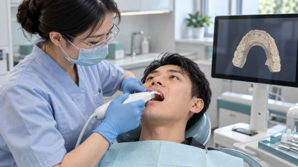
Integrating facial scans and intraoral scans means positioning detailed dental geometry within a three-dimensional facial reference, checking that spatial relationship, and using it to guide veneer shape, position, and clinical evaluation. The central task is registration: establishing where the teeth actually sit relative to the face.
I judge Digital Smile Design by the decisions its records can support. A convincing image has value. But I would require separate evidence for tooth position, occlusion, preparation geometry, and the design eventually released for fabrication.
What Each Scan Contributes to Digital Smile Design
A 3D virtual patient is an assembled model. Its reliability depends on the source records and the transformations used to combine them.
Before opening the design software, I would define the intended changes using a brief such as the five esthetic goals to establish before veneer design. Otherwise, the team can spend an afternoon perfecting a model without agreeing on the desired incisal position.
Here is how I would assign responsibility to each record.
Record
Main contribution
Review before using it
Facial scan at rest
Facial surface, lip position, and overall orientation
Expression, head position, incomplete surfaces, and distortion
Facial scan during smiling
Tooth display and the surrounding lip frame at that captured moment
Smile consistency and usable dental references
Preoperative intraoral scan
Existing tooth anatomy and a baseline for proposed changes
Missing surfaces, duplicated geometry, and scan completeness
Opposing arch and bite records
Relationship between the upper and lower dentition
Agreement with the clinician’s recorded occlusion
Preparation scan
Geometry needed to define the restoration’s fit and margins
Visible finish lines, tissue interference, and surface defects
Approved mock-up or provisional scan
The physical design accepted after clinical evaluation
Version, documented modifications, and correspondence with the approval record
These records answer different questions. A smile scan shows one expression. A bite record captures one jaw relationship. We should be explicit about what each dataset represents before treating the combined model as a complete account of the patient.
How to Integrate Facial Scans and Intraoral Scans for Veneer Design
1. Capture a coherent set of clinical records
For intraoral scanning for veneers, I would request the preoperative dentition, opposing arch, and an appropriate bite record, followed by preparation scans when that stage is reached. The clinician should identify any intended change in the recorded jaw relationship.
For the facial records, capture rest and smile conditions separately. Label them clearly. Include photographs or short video showing the expressions and speech the clinician wants the laboratory to understand.
A retracted registration record also needs a clear label. Retraction changes the visible lip frame, so it should not quietly become the reference for the patient’s natural smile.
The clinic and laboratory can organize these materials around the anterior veneer case submission checklist. For scan integration, I would add the acquisition date, device, software version, facial expression, and registration method to that package.
Small omissions travel.
If the technician cannot distinguish a preoperative scan from an approved provisional, every subsequent comparison becomes questionable.
2. Confirm file compatibility before the handoff
Check what the receiving software can import and what the scanner actually exports.
For example, exocad’s mesh-import documentation lists STL, OBJ, PLY, and other supported mesh formats, with separate roles for face scans, preoperative models, and bite alignment scans. That is a useful starting point for a compatibility discussion.
I would ask for:
Original exported meshes, with their intended units documented.
Any accompanying texture images or material files produced by the export.
Separate identification of upper arch, lower arch, bite, face, and approved design.
The native project when needed to preserve alignment or case information.
Confirmation that the receiving system opens the files correctly.
Keep the dental geometry intact. If an imported model appears at the wrong scale, investigate the export and unit settings before making a visual adjustment.
3. Establish the dental-to-facial relationship
Facial and intraoral scan integration needs recognizable correspondence between the datasets.
Where sufficient dental surfaces are captured in both scans, those surfaces can help establish the relationship. Where overlap is poor, I would request additional acquisition through the chosen system’s registration protocol before proceeding.
The software workflow matters. In exocad’s Smile Creator instructions, the face-scan sequence begins with two corresponding registration points and then permits manual adjustment of the jaw relative to the face, including inspection from different orientations.
Those two points begin that particular workflow. They do not independently certify the finished alignment.
My review would preserve the detailed intraoral geometry while checking the positioning operation. Any unexplained scaling or deformation of the dental mesh would need resolution before design approval.
4. Inspect the registration from several directions
Rotate the model.
A frontal view can conceal depth discrepancies that become obvious from the side. I would inspect the registered teeth from frontal, lateral, and oblique views, then compare recognizable dental features across the overlapping records.
The review questions are practical:
Do corresponding incisal edges occupy the same position?
Is there visible rotation or a depth offset across the anterior segment?
Are duplicated contours present?
Does an apparent match depend on distorted or incomplete scan regions?
Does the imported bite still agree with the clinical record?
If the alignment is repeated, does the reviewer obtain a similar result?
Where the software provides a deviation map, record the measured region, units, and metric. A whole-face average and a local dental discrepancy describe different things.
I would also retain the alignment views used for approval. A reviewer should be able to see why the team accepted the registration.
5. Design the veneers against a documented facial target
Once the spatial relationship is accepted, digital veneer design can address tooth length, apparent width, incisal position, midline, embrasures, and the visible transition toward the canines.
I would separate three decisions in the case notes: what the patient wants to change, what the clinician considers appropriate, and what the laboratory is authorized to modify.
That distinction becomes useful when a requested increase in visible tooth length affects the proposed contour or functional relationships. The technician needs an explicit decision to work from.
6. Evaluate the proposed design clinically
The digital design should lead to a clinical evaluation of an appropriate mock-up or provisional.
For that review, I would request documentation of appearance at rest and during smiling, speech, lip support, and the clinician’s assessment of occlusion. Record any changes made during the appointment.
Then scan the accepted result.
The final CAD reference should identify those accepted modifications. If the patient approved a shortened incisal edge chairside, the laboratory needs that updated geometry and a clear revision record.
What Three Published Studies Actually Show
The research supports useful applications of scan integration. It also gives us reasons to keep accuracy claims specific.
A 2018 clinical report demonstrated a veneer application
In their clinical report on facial scanning and CAD-CAM ceramic veneers, Wei-Shao Lin and colleagues described a workflow combining virtual smile design with a static, photorealistic 3D patient to restore maxillary central incisors using monolithic lithium disilicate veneers.
The report appeared in the February 2018 issue of the Journal of Prosthetic Dentistry, following online publication in June 2017.
This is directly relevant evidence of clinical feasibility. A case report cannot establish a general reduction in remakes, improved long-term survival, or superiority across scanner systems.
A 2024 study measured automation speed and regional mismatch
Reported average registration times were 26.5 seconds for the AI-driven approach, 30.8 seconds after AI refinement, and 385 seconds for the semiautomatic reference workflow. The greatest facial-scan-to-CT mismatch occurred in the lower facial third.
My reading is that faster assembly still requires regional inspection. This was a multimodal registration study; it did not demonstrate a veneer survival benefit or establish a need for CT in routine veneer planning.
A 2025 smartphone study shows why the measurement matters
They reported a mean three-dimensional root mean square deviation of 1.47 mm, a mean absolute linear difference of 1.43 mm, and a mean angular difference of 3.16°. Participants already required CBCT as part of their care.
Those are facial comparison measurements. They cannot be used as veneer marginal-gap measurements or as a universal acceptance threshold for dental-to-facial registration.
For me, the purchasing question follows naturally: which task was validated, against what reference, and in which region?
CAD/CAM Veneer Design Needs a Manufacturing Brief
An accepted facial arrangement still leaves manufacturing decisions to resolve.
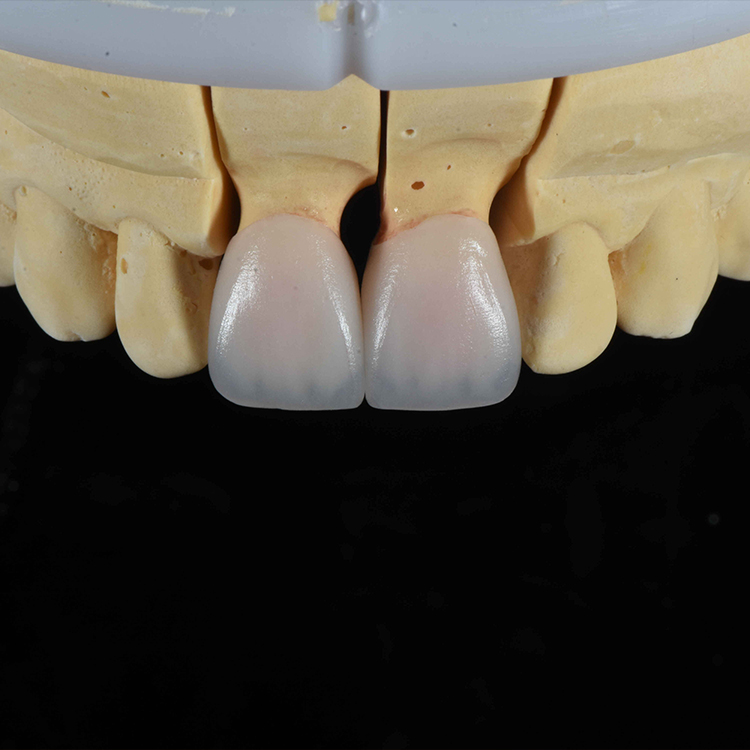
For lithium disilicate E.max veneers, the laboratory needs preparation and bite information alongside shade, stump shade, photographs, and design instructions. The material page’s requested records make the handoff concrete.
I would keep the production brief focused on the decisions that affect the prescribed restoration:
The exact ceramic system and processing route.
Margin location and preparation design.
Available restorative space and material-specific thickness requirements.
Contact and occlusal instructions.
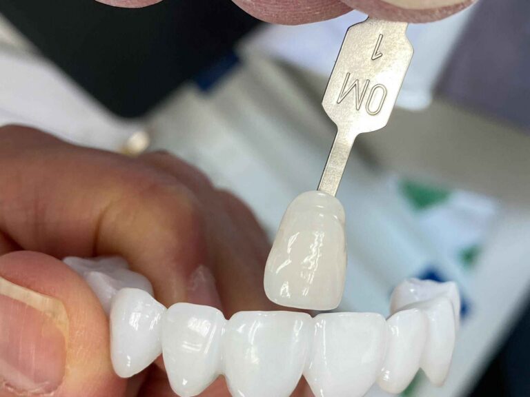
Final shade, stump shade, translucency, texture, and surface finish.
The approved design version and permitted modifications.
“E.max” alone does not specify every production choice. Ask the laboratory to identify the intended product and fabrication route, then coordinate those choices with the clinician’s prescription and the applicable manufacturer instructions.
And keep digital appearance in perspective. A rendering should not be treated as a calibrated prediction of the final ceramic shade.
The Approval Records I Would Require Before Production
My proposed control is simple: retain a short record of what was approved at each stage.
First, approve the incoming data and registration. Next, approve the proposed design after clinical evaluation. Finally, approve the manufacturing parameters against the current prescription.
For this integration task, I would retain five items:
The original scans and acquisition details.
The registered project and the views used to inspect it.
The proposed design with its revision identifier.
The clinically approved mock-up or provisional record.
The released CAD version and laboratory prescription.
We can then trace a change in incisal position to a specific decision. Without that record, a disagreement about the delivered result can become an argument over which screenshot everyone remembers.
FAQs
What is Digital Smile Design for veneers?
Digital Smile Design for veneers is a planning and communication process that uses facial records, dental scans, and proposed tooth forms to evaluate the intended smile, coordinate clinical and laboratory decisions, and guide an approved design toward a mock-up, provisional, or definitive restoration.
Its usefulness depends on the records and the clinical evaluation supporting those decisions.
How do you integrate facial scans and intraoral scans for veneer design?
Integrating facial scans and intraoral scans for veneer design involves importing compatible datasets, establishing correspondence between their shared references, aligning the dental model within the facial record, inspecting that registration, and transferring the proposed tooth arrangement into a mock-up or provisional for clinical evaluation.
The specific alignment sequence depends on the software and acquisition protocol.
What is a 3D virtual patient in dentistry?
A 3D virtual patient is a composite digital representation that brings dental and facial records into a shared spatial framework, allowing clinicians and technicians to examine anatomical relationships and proposed restorations while retaining the limitations of each source dataset and its registration method.
Additional imaging or movement records may be incorporated when appropriate to the clinical task.
Can a smartphone facial scan be used for veneer planning?
A smartphone facial scan can contribute facial context to veneer planning when the particular device, application, acquisition conditions, and registration method provide suitable data for the intended task, while dental detail, preparation margins, and occlusal relationships remain dependent on their respective clinical records and verification.
Confirm the complete workflow before relying on a particular device.
How accurate must facial and intraoral scan integration be?
The required accuracy of facial and intraoral scan integration depends on the intended clinical decision, the anatomical region being assessed, and the validated acquisition and registration method, so acceptance should consider local discrepancies and clinical verification alongside any reported overall surface deviation metric.
The studies discussed above do not establish one universal acceptance threshold for veneer cases.
What should the laboratory receive for a digital veneer workflow?
For a digital veneer workflow, the laboratory should receive clearly identified dental and facial records, an appropriate bite record, preparation data when available, shade information, the proposed or approved design, relevant clinical instructions, and documented revisions explaining which geometry and esthetic decisions are ready for fabrication.
Include file formats and software details so compatibility can be checked before design begins.
Request a Review of Your Next Veneer Case
Before starting your next case, identify the records you can provide and the design decisions that still need agreement.
Request a digital veneer case review from Artist Dental Lab and include your scanner and software names, available file formats, proposed restorations, and current design or mock-up status. Ask for confirmation of file compatibility, any missing records, the intended material route, and the approval steps before fabrication.
Agree on the file-transfer method, then submit the case package with one clearly identified current version.